Thursday, October 4, 2012

It's a bee - no, it's a fly - no, it's a BEE FLY!


The biology professors here at Eastfield College often take their students into the field.  Professor Ron Beecham took a group of students to Gus Engeling Wildlife Management Area last weekend and brought some samples back to the lab for me to image.  One of these is a bee fly - genus Bombylius.

This little guy is easily recognizable as a fly because it possess a single pair of wings and a pair of halteres - reduced wing stubs that are thought to act as counterbalances.  In fact, the order for flies is diptera, which literally means "two wings". ("di-" is pair and "ptera" is wing.)

 Dissecting Scope Image - Ventral View
Note the long tongue

 Dissecting Scope Image - Ventral View
The haltere on the left side of the fly appears as a yellowish blob due to the limited depth of field of the camera on the scope.

Dissecting Scope Image
Dorsal View

As I examined this guy, I noticed a tuft of some "hairs" on the leading edge of the wing near the body.  This tuft if visible in the following two images.

Dissecting Scope Image
Tuft of hairs on right wing, near body.
Dissecting Scope Image
A closer look at tufts of hairs on right wing.

Now for a head-on view of the fly.  The multiple facets of the ommatidia that make up the compound eye are obvious, as are the antennae.

Dissecting Scope Image

Now on to the Scanning Electron Microscope.  Here is pretty much the same pose and you can see the advantages, and single disadvantage, of scanning electron microscopy.  The only disadvantage with the SEM is loss of color since we are using a beam of electrons instead of light.  Not much of a disadvantage at all as far as I am concerned.  One big advantage, which is very obvious when comparing the image above with the one below, is the depth of field.  On the top image I focused on the upper surface of the eyes - the rest of the image is out of focus.  Below you can see that much more of the specimen is in focus.

Head shot
25x


 Head shot
37x
My, what beautiful eyes you have!
 Right eye
85x

Ommatidia of the compound eye - 20 micros across
267x

Antenna
Note the pores
160x

The next series of images are of the haltere, or reduced second pair of wings.


 Haltere
60x


Close-up of haltere - everything has structure
170x

(I realize as I am labeling this image that I missed an opportunity.  Is the structure on the haltere similar to that on the wings of the insect?  A question needing an answer - sounds like an opportunity for someone to do some science.)

As you may have noticed, this little fly has a very long tongue.  Let's see what it looks like

 Tongue
80x
At a higher magnification you can see some pollen grains that are stuck to the tongue.
98x
 Pollen grains and tongue structure
From the scale bar you can see that the pollen grains are between 30 and 35 microns in diameter.
394x

 Tip of the tongue
145x

 Tip of the tongue
447x

 As I was imaging the tongue I noticed some interesting structures on the legs of the fly.  They appear to be covered with scales very similar to those found on butterflies.  This may be normal, but being new to SEM work, it definitely surprised me.
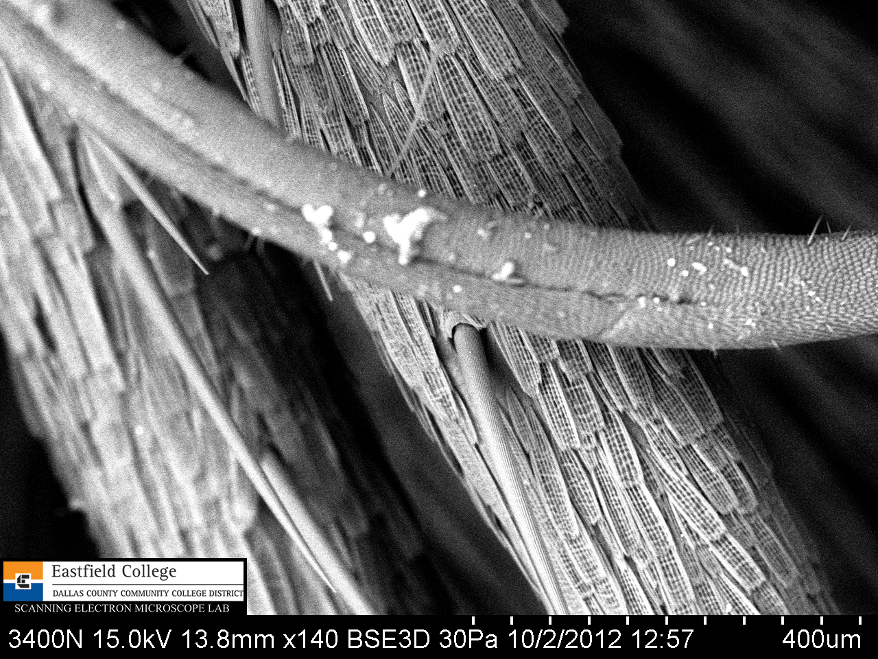

 Tongue in foreground, leg with scales behind
140x
Leg with spines
50x
 Leg joint showing scales and fine structure of spines
190x
Science is all about making observations, asking questions, and then finding out the answers to those questions.  What is the function of scales - on flies and butterflies?  Is it possible they have some aerodynamic effect?
Now a look at the wings. 


 Wing
Note the "hairy" structure on the leading edge of the wing at the top of the image.
27x
 The tufts of "hair" are actually spines and scales.
65x
Scales and spines on leading edge of the wing.
182x


Eastfield College makes it Scanning Electron Microscope lab available to students and faculty at all levels.  If you would like to see or use our SEMs (we have two) please contact me.  Our smaller SEM is portable and can be brought to your school for students to use.  I know, it sounds like a sales pitch, but for high schools, middle schools, and elementary schools there is not cost.

Our lab is also open for visits - bring your classes.  I also Skype!

Murry Gans
Scanning Electron Microscope Lab Coordinator
Eastfield College
Mesquite, TX


Thursday, September 13, 2012

Face to Face with a Brown Recluse


Biologist have something interesting happen to them all the time - people bring them organisms out of the blue.  I cannot tell you how many times I have been presented with or pointed toward some sort of creature.  Snake in the building - get the biology teacher.  Dead bird on the sidewalk - get the biology teacher.  Find a dessicated gecko under a carpet tile? Balance its stiff body on the door handle of the lab - you just know he wants to see it. 

It sounds like I am complaining, but I have gotten some really cool stuff this way.  Just last week one of my colleagues here at Eastfield walked into my lab with a rather large Brown Recluse  spider (Loxosceles reclusa) in a jelly jar.  He knows I like spiders and, being the true biology nerd that I am, it was pretty cool.

Not only was this particular spider a Brown Recluse, but it was a honking big, mature male.


 Dorsal Portrait
The characteristic "violin" marking on the cephalothorax is very obvious. 
Note also the two large pedipalps in front of the chelicerae.

 In this close up you can see that the "violin" marking is due to a dense patch of hairs. 
Notice also that this is a 6-eyed spider. 

Ventral view with a focus on the pedipalps - the male sex organs. 
That long spike, or embolus, fits specifically into the genital opening of the female.

[31x] 
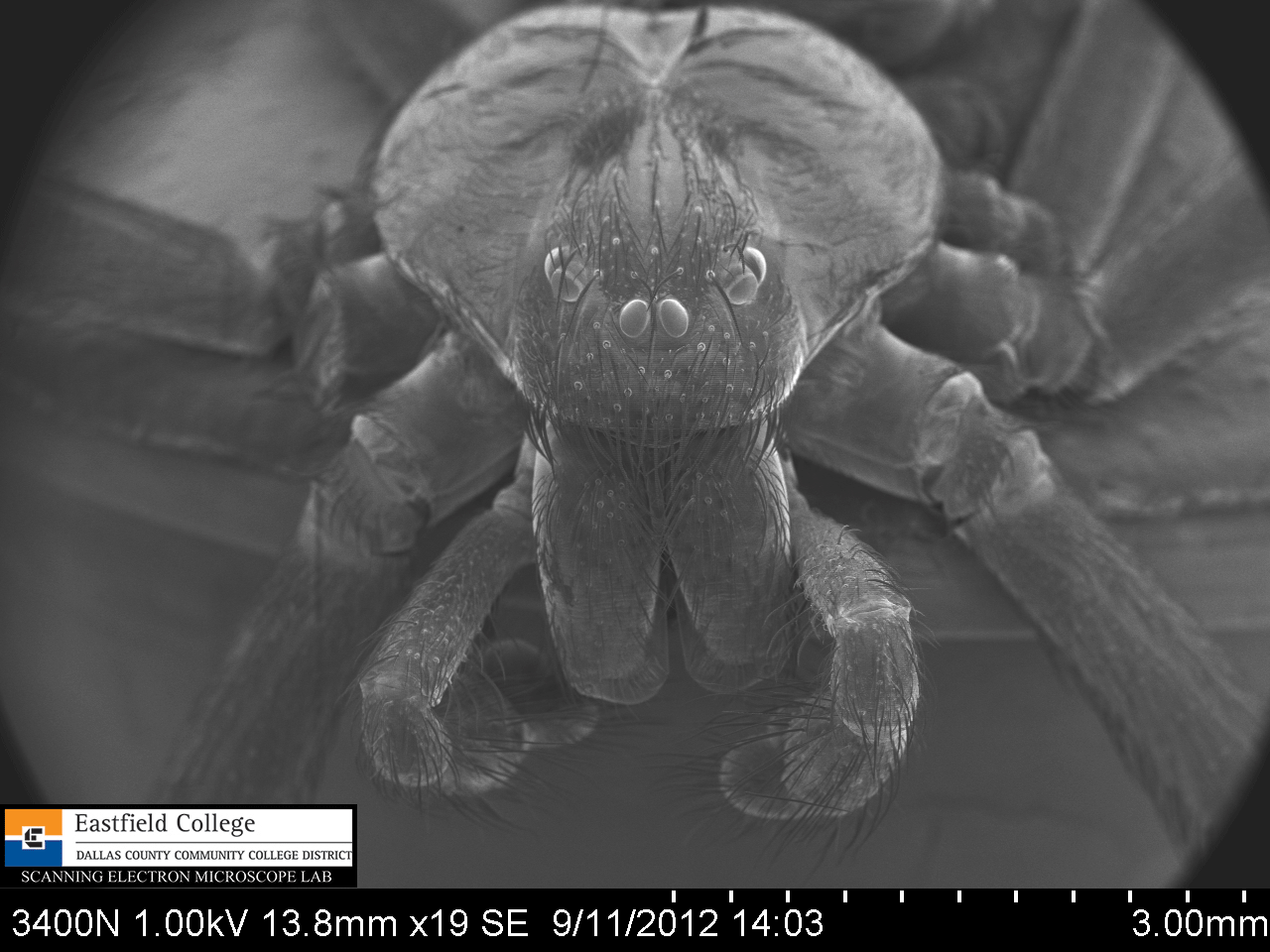
Hi there!  Face to face with a spider that you should be afraid of.
This image shows two main characteristics for identification - three pairs (dyads) of eyes in a backwards facing curve (strongly recurved) and fused chelicerae.

[19x]
This image is less magnified and more dorsal.
Recurved eye dyads

[93x]
A close up of the eye dyads.
I didn't intend it to be, but this is kind of a creepy picture.

[18x]
Ventral view showing mouth parts (labium and endites), chelicerae, cheliceral fangs, and pedipalps with bulbs and embloli.

 [42x]
Left pedipalp with bulb and embolus


 54x
Right pedipalp with bulb and embolus

 [99x]
Close up of bulb

 [70x]
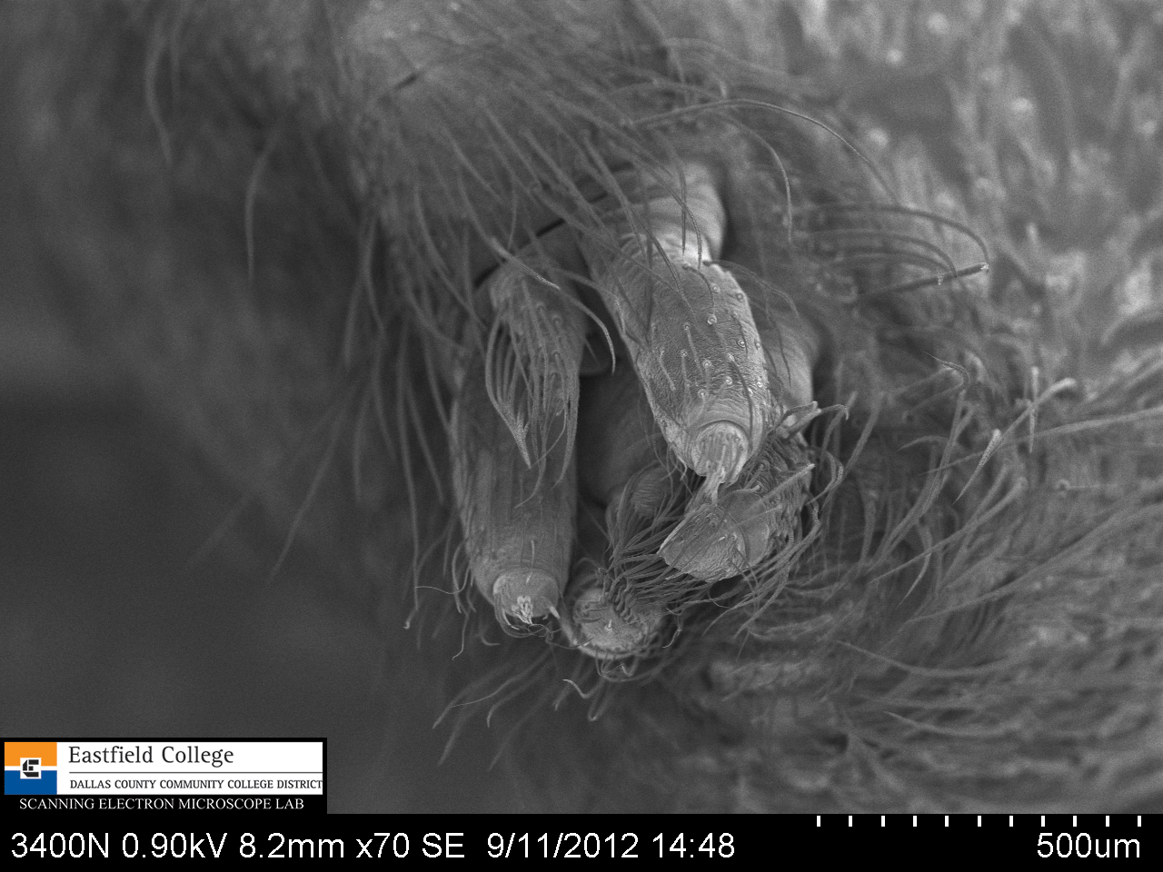
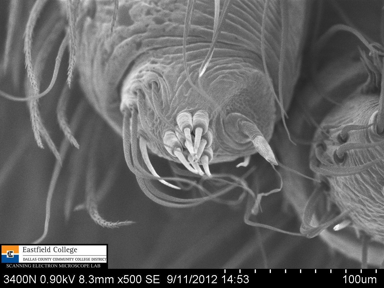
Spinnerets

 [320x]
Spinnerets

[500x]
Spinnerets

[82x]
I mounted the spider on its back to get a clear view of the cheliceral fangs.
In the image above the openings through which the spider injects its venom are clearly visible.

 [170x]
A close up of one of the fangs.

 [321x]
I will admit that I am very pleased with this image.  This extreme close up of the cheliceral fang not only shows the opening for injecting venom but also the serrated edge of the fang.

This spider turned out to be fairly hard to work with - not because it is a poisonous spider, but because it has unusually long legs.  Positioning him for images was difficult and resulted in me accidentally piercing his abdomen.  Drat!  No worries though, I am sure someone will bring me one of his relatives any day now.

If you would like to begin to make your own discoveries please remember that Eastfield College supports student and faculty research at all levels - in fact I am talking to some 3rd, 4th, and 5th graders next week.
You are invited to come to Eastfield to use our scopes.  If you are not in the area, I would be delighted to Skype with classes.

Hope to hear from you soon!

Murry Gans
Scanning Electron Microscope Lab Coordinator
Eastfield College
Mesquite, TX
972-860-7267


Rosemary's Spider


Eastfield College has some beautiful courtyards with lots of shade and benches.  Even in our extremely hot Texas summers I can always find a place to sit and relax for a few minutes.

The other day I was sitting on one of my favorite benches when I noticed that the rosemary in the planter next to me was covered in spider webs.  I am a good enough arachnologist to realize that where there are spider webs there are spiders.  I took a quick look but couldn't find the little creatures so I clipped off a sprig of rosemary and took it back to my scopes in the SEM Lab.


Once I got the sprig of rosemary back to the lab I put it on the dissecting scope and began my search.  Within a few minutes I found the little guy.


Another very small Theriidian spider.  (You can see another species of this same family of spiders in a previous blog.)  These spiders are commonly referred to as cobweb spiders. Black widow spiders belong to the same family.

By this time you can imagine that my lab and hands are smelling very strongly of rosemary.  All of that aromatic oil must be stored in glands on the leaf.

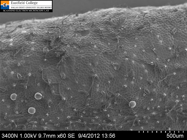
A look at the top surface of the leaf shows the expected oil glands.  If you look carefully at the image above you can see there are two different sizes of glands.  Do they contain different oils? Sounds like a good question for someone with a gas chromatograph.

As observant as I try to be I am a bit embarrassed to admit that I had never looked closely at the leaf on the rosemary bush - and I have one in my own backyard. 

What I didn't realize is that the top surface of the leaf curls around to the bottom of the leaf.


 This image shows the curled edges of the top surface of the leaf out of focus at the top and bottom.  The brighter out of focus structure in the middle is the midrib of the leave.  Even at this magnification you can clearly see the densely packed oil glands.

 A closer look at the oil glands on the lower surface of the rosemary leaf.

The images above illustrates a problem - a limited depth of field.  How to solve that problem - don't use light - use electrons in the SEM.


 Top surface of rosemary leaf. [60x]
The image above shows the epidermal cells of the leaf as well as the two different sizes of oil glands.

 Lower surface of the rosemary leaf [24x]
Note how all levels of the leaf are now in focus.

 Lower surface of rosemary leaf [70x]
This image shows the midrib with smaller oil glands.  You can also see some spider web stretching diagonally across the upper half of the picture.

 Spider web [99x]

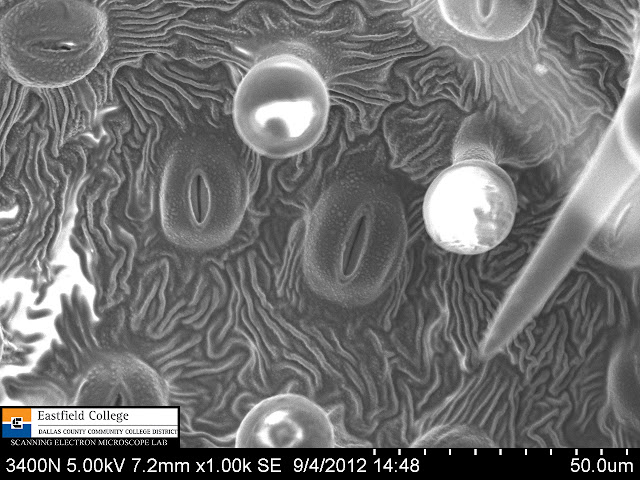
 Stomata and oil glands [1,000x)

 Stoma portrait [2,490x]
The high vacuum and electron beam of the SEM dry out specimens.  This stoma was open when I first saw it, but quickly closed as the guard cells lost their turgor.

As I use the instruments in the SEM Lab at Eastfield College I am discovering that even the most common organisms have some pretty fantastic morphology under high magnification.


The imaging instruments at Eastfield College are available for research to students and faculty everywhere.  Want to get a better look at your world - give us a call.

Murry Gans
Scanning Electron Microscope Lab Coordinator
Eastfield College
Mesquite, TX
972-860-7267