Biologist have something interesting happen to them all the time - people bring them organisms out of the blue. I cannot tell you how many times I have been presented with or pointed toward some sort of creature. Snake in the building - get the biology teacher. Dead bird on the sidewalk - get the biology teacher. Find a dessicated gecko under a carpet tile? Balance its stiff body on the door handle of the lab - you just know he wants to see it.
It sounds like I am complaining, but I have gotten some really cool stuff this way. Just last week one of my colleagues here at Eastfield walked into my lab with a rather large Brown Recluse spider (Loxosceles reclusa) in a jelly jar. He knows I like spiders and, being the true biology nerd that I am, it was pretty cool.
Not only was this particular spider a Brown Recluse, but it was a honking big, mature male.

Dorsal Portrait
The characteristic "violin" marking on the cephalothorax is very obvious.
Note also the two large pedipalps in front of the chelicerae.

In this close up you can see that the "violin" marking is due to a dense patch of hairs.
Notice also that this is a 6-eyed spider.

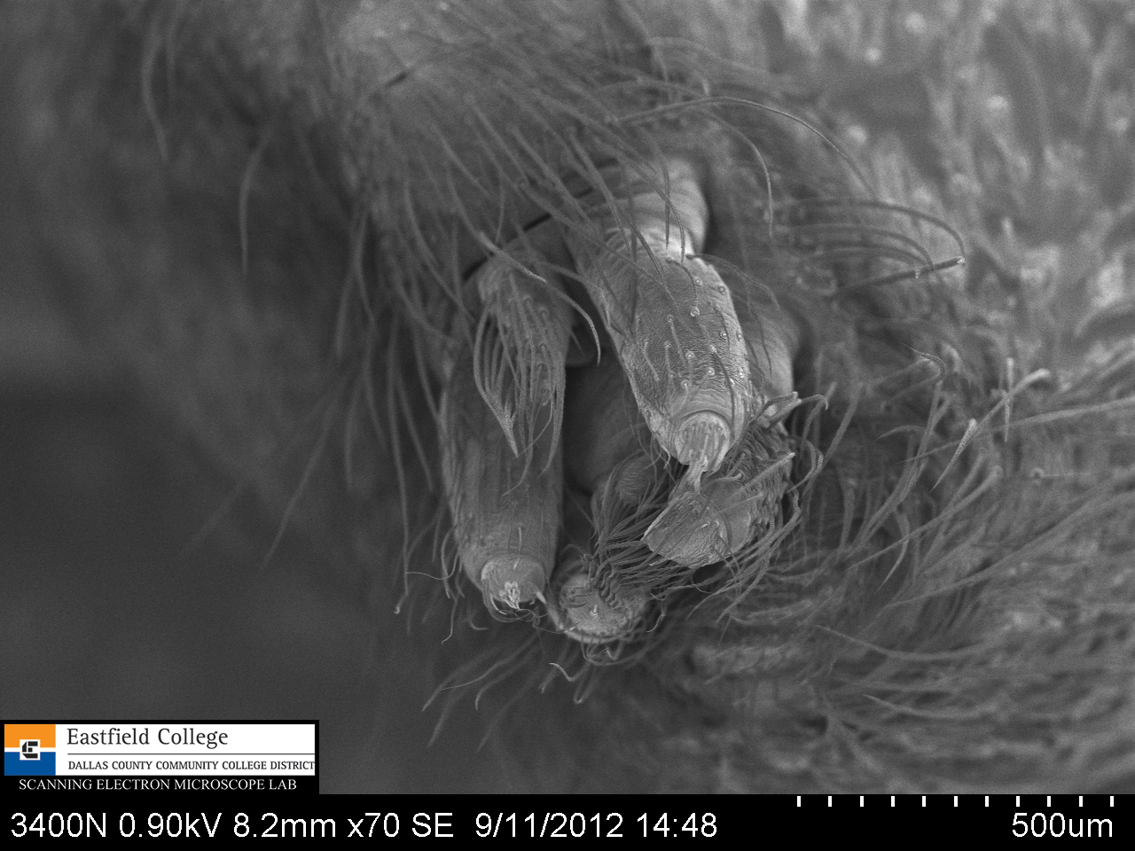
Ventral view with a focus on the pedipalps - the male sex organs.
That long spike, or embolus, fits specifically into the genital opening of the female.

[31x]
Hi there! Face to face with a spider that you should be afraid of.
This image shows two main characteristics for identification - three pairs (dyads) of eyes in a backwards facing curve (strongly recurved) and fused chelicerae.
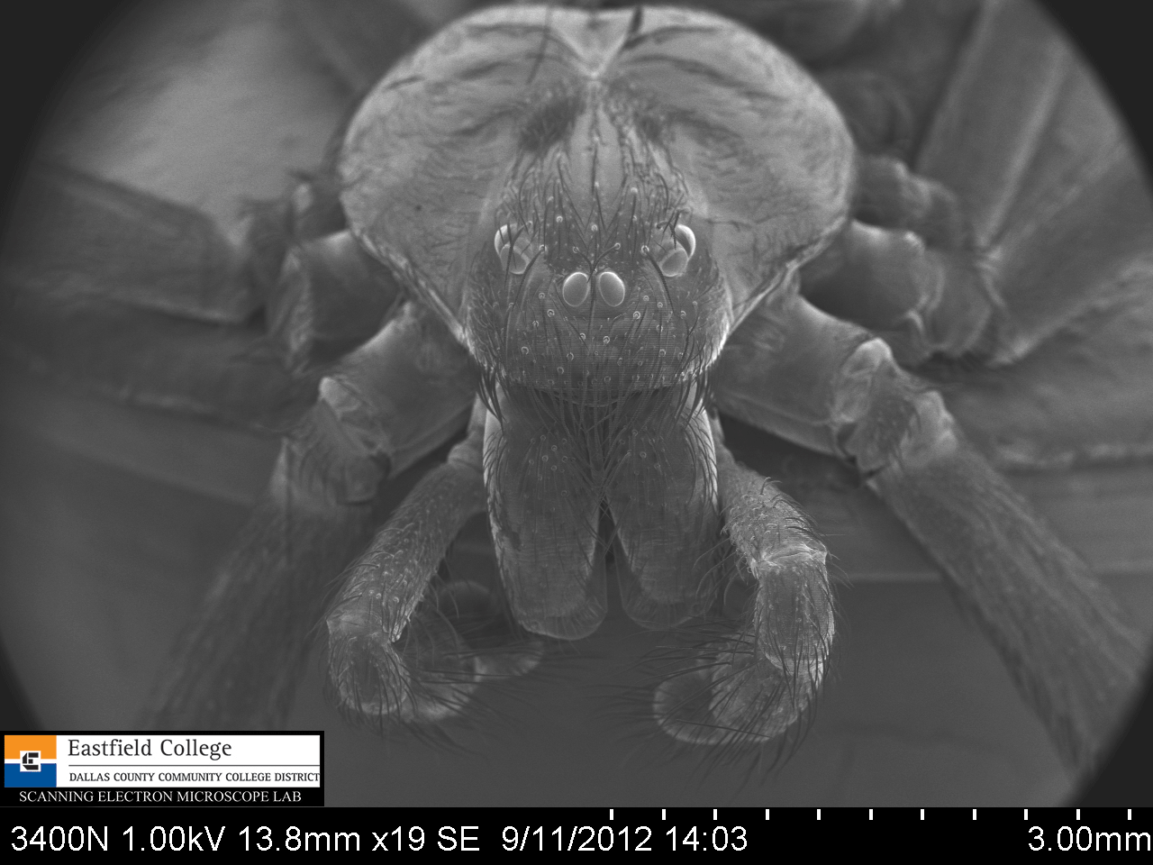
[19x]
This image is less magnified and more dorsal.
Recurved eye dyads

[93x]
A close up of the eye dyads.
I didn't intend it to be, but this is kind of a creepy picture.

[18x]
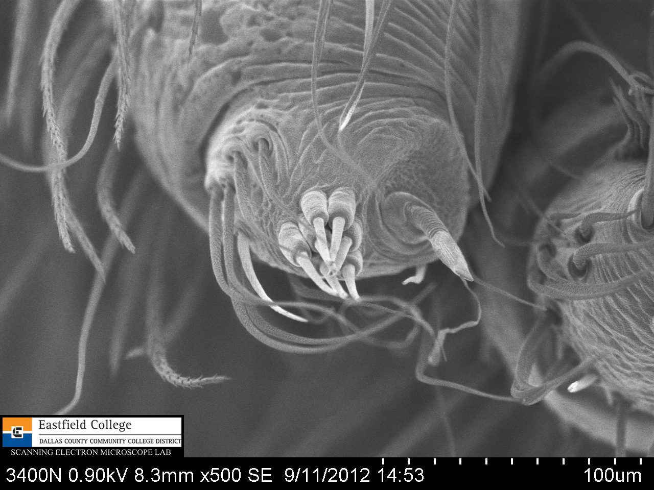
Ventral view showing mouth parts (labium and endites), chelicerae, cheliceral fangs, and pedipalps with bulbs and embloli.

[42x]
Left pedipalp with bulb and embolus

54x
Right pedipalp with bulb and embolus

[99x]
Close up of bulb
[70x]
Spinnerets

[320x]
Spinnerets
[500x]
Spinnerets

[82x]
I mounted the spider on its back to get a clear view of the cheliceral fangs.
In the image above the openings through which the spider injects its venom are clearly visible.

[170x]
A close up of one of the fangs.

[321x]
I will admit that I am very pleased with this image. This extreme close up of the cheliceral fang not only shows the opening for injecting venom but also the serrated edge of the fang.
This spider turned out to be fairly hard to work with - not because it is a poisonous spider, but because it has unusually long legs. Positioning him for images was difficult and resulted in me accidentally piercing his abdomen. Drat! No worries though, I am sure someone will bring me one of his relatives any day now.
If you would like to begin to make your own discoveries please remember that Eastfield College supports student and faculty research at all levels - in fact I am talking to some 3rd, 4th, and 5th graders next week.
You are invited to come to Eastfield to use our scopes. If you are not in the area, I would be delighted to Skype with classes.
Hope to hear from you soon!
Murry Gans
Scanning Electron Microscope Lab Coordinator
Eastfield College
Mesquite, TX
972-860-7267

No comments:
Post a Comment