Tuesday, August 21, 2012

Itsy Bitsy Spider - The Scanning Electron Microscope Version


Even if you aren't interested in the biology of spiders, please check out the images in this blog posting.  These small and often loathed creatures have some of the most remarkable anatomy you will find on the planet, and they are in the most mundane places - like around the porch light in my backyard.

In my previous blog (August 20, 2012) I showed images of a small Theridiid - a cobweb spider.  The images in this posting were made with the scanning electron microscope here at Eastfield College.
 
Prosoma (Dorsal)
70x
A shot of the dorsal surface of the cephalothorax or prosoma.  Even at 70x you can immediately see that this spider does indeed have 8 eyes, not just the 6 I thought I saw with the light dissecting scope.
 
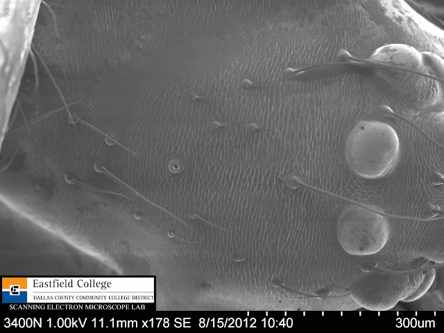
Prosoma
178x
Several hairs on the prosoma and one obvious sensory pit.

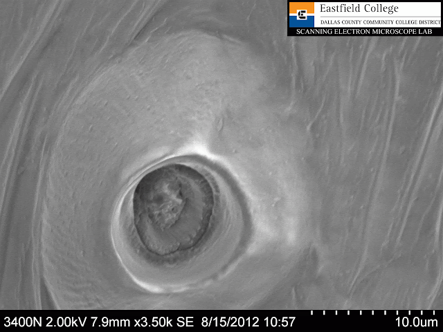
Sensory Pit
3,500x
Small sensory pit on dorsal surface of prosoma.  Distance between marks on scale = 1 micron.
 
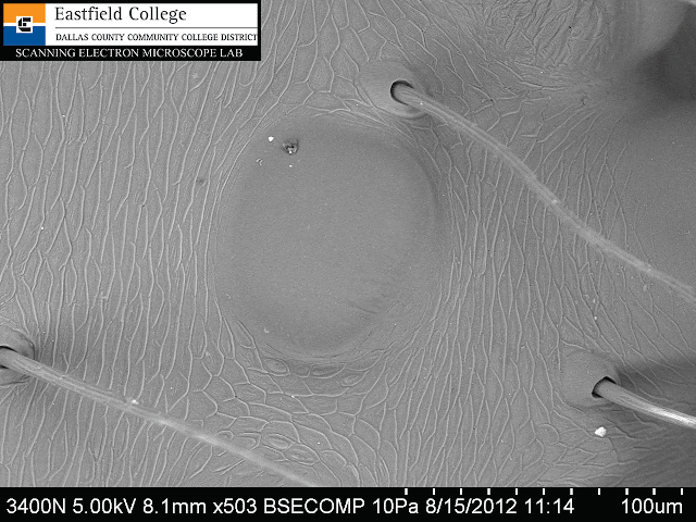
PME
503x
Spiders do not have compound eyes like insects.  This image is of the PME or median eye on the posterior eye row.
 
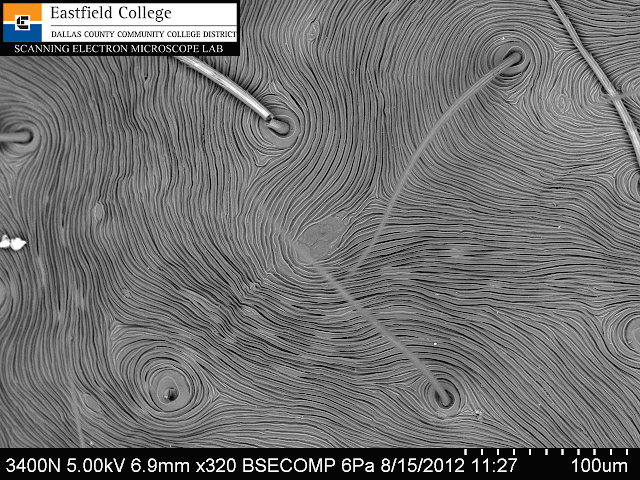
Dorsal Opisthosoma
320x
Image of the peak of the opisthosoma or abdomen.
 
Anterior Opisthosoma
190x
To make this image I rotated the spider so he was facing me - the bright structure at the bottom of the image is the prosoma.  Basically I was shooting electrons right over his head and onto the abdomen.


Side portrait
47x

Pedicel
250x
This is a side shot of the spider focusing in on the connection, or pedicel, between the two body parts.  In this image the spider is facing to the left.  Prosoma to the left, opisthosoma to the right.

Prosoma or cephalothorax
90x
The dyad of eyes (pairing) is very obvious in the micrograph.  The dyad is composed of the ALE and the PLE - the lateral eye of the anterior eye row and the lateral eye of the posterior eye row.

 
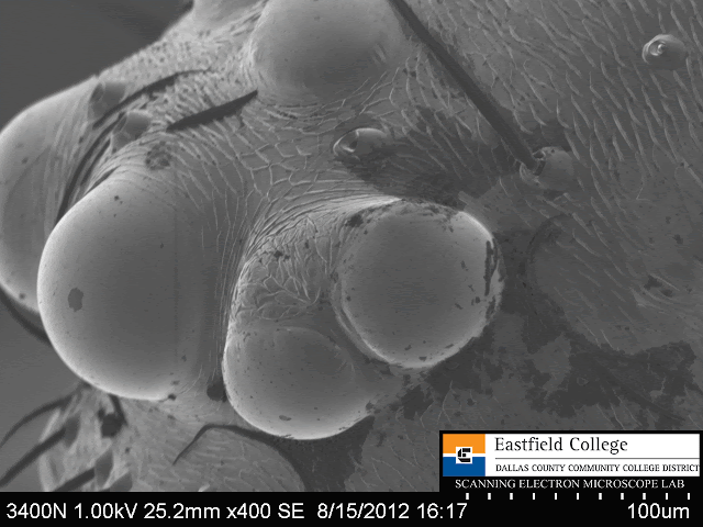
ALE/PLE Dyad
400x
The dyad is composed of the ALE and the PLE - the lateral eye of the anterior eye row and the lateral eye of the posterior eye row.

Ventral Abdomen - Epigastric Furrow
60x
  
Epigastric furrow and epigynum
300x
Ventral view of anterior region of abdomen of opisthosoma.  The large fold across the photo is the epigastric furrow and the opening with the concentric rings around it is the epigynum, or reproductive opening of the female.
  
Epigynum
804x

Spinnerets
200x
There are three pairs of spinnerets.  Most obvious are the large, paired anterior spinnerets at the top of the image and the large, paired posterior lateral spinnerets just below them.  (The football shaped structure at the bottom of the spinnerets is the anus.)  Not so obvious are the smaller, paired posterior median spinnerets between the larger PLS.
 
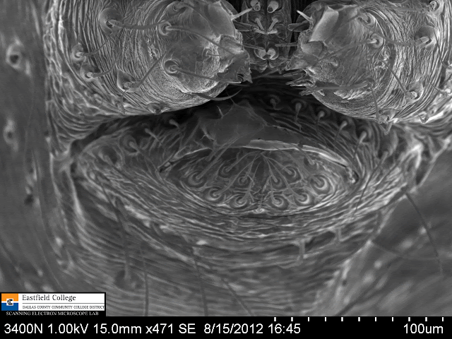
Anus and Posterior Spinnerets
471x
Posterior lateral and median spinnerets at top of image.
  
Close up of Median Posterior Spinnerets
1,090x

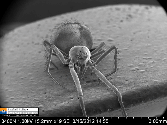
Portrait
19x
I wanted to image the spider head on so I draped its front pair of legs over the mount.

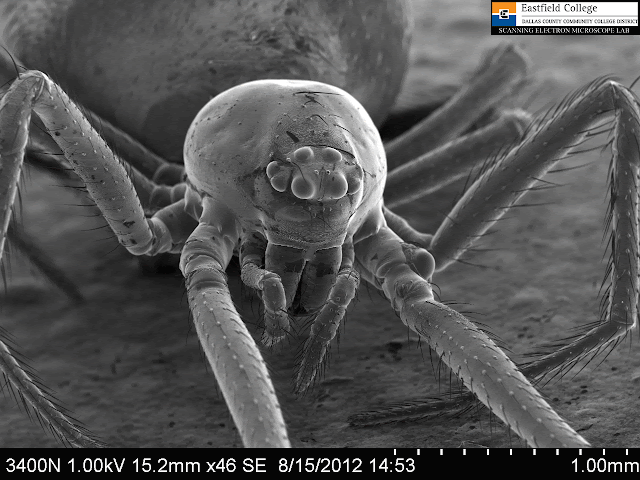
Theridiid Head Shot
46x


Eye Arrangement
230x
Image of the two row of eyes on this spider - the anterior eye row (AER) and the posterior eye row (PER).

Close up of Posterior Median Eyes (PME)
470x
Note the sensory pits above the eyes.
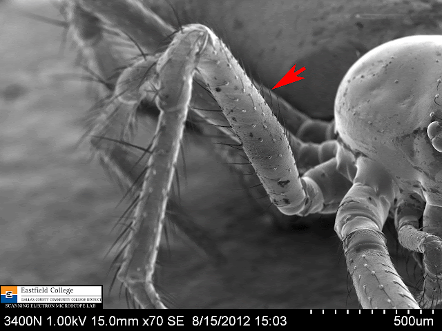
Now for some basic spider leg anatomy! 
This study focuses on the second leg (simply because it was in the proper position).
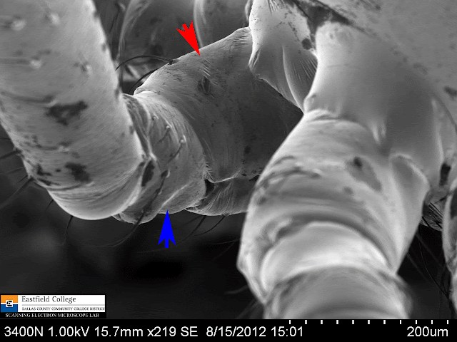
Coxa and Trochanter
219x
The coxa is indicated by the red arrow and the trochanter by the blue arrow.


Femur
70x

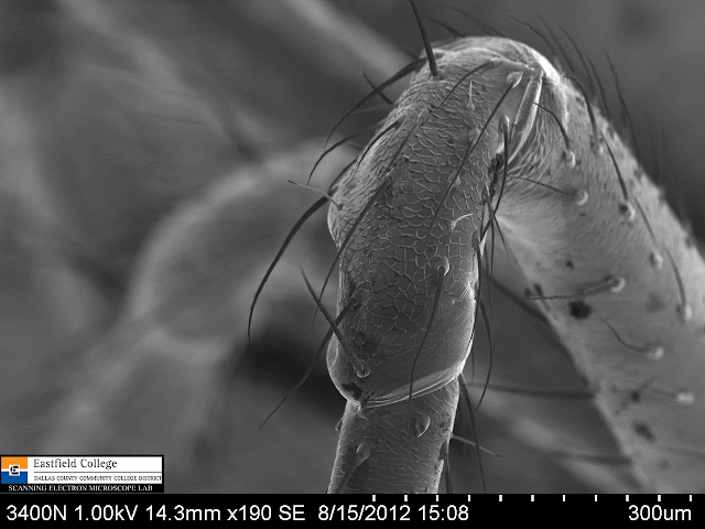
Patella (Knee)
190x
 
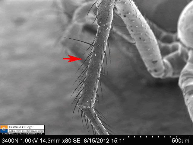
Tibia
80x

Close up of hairs on tibia
422x

(Drat!!  I just figured out that I became fixated on the hairs on the tibia and didn't image the last two segments of the spider leg, the metatarsus and tarsus.)


Egg Sac
30x
In this image the egg sac has been teased open showing the spiderlings inside.  In the upper right of the photo is a spiderling who was outside of the egg sac.

[Technical note:  The difference in brightness of the top and bottom parts of this image is caused by the specimen accumulating a charge.  Spiderlings at the top of the photo were not in contact with the mounting so they became negatively charged, which causes them to glow.  Not what I would have liked, but still an interesting image]


Spiderling Portrait
120x
Distance between marks on scale = 40 microns.  This guy is little!
 
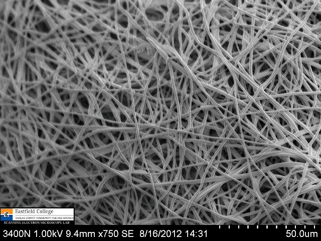
Egg Sac Silk
750x
 
Egg Sac Silk
2,100x
Diameter of silk strands less than 2 microns

After two days of imaging and a couple of days of selecting images for the last two blogs, I can truthfully say that I have totally satisfied my spider jones . . .  for now. 

Eastfield College is more than happy for you to use our scanning electron microscopes to satisfy your own curiosity whether that be biology, physics, or chemistry.  We provide the SEMs, you provide the specimens.  All you have to do is contact us.

Murry Gans
Scanning Electron Microscope Lab Coordinator
Eastfield College
Mesquite, TX
972-860-7267


Monday, August 20, 2012

Itsy Bitsy (Cobweb) Spiders - Thank goodness for good tools!


For many years I have been intrigued with the very small spiders that live around the porch light and back door frame at my home in Irving, TX.  Now that I have the SEM Lab at Eastfield, I can finally get a good look at them and classify them.

In fact, I may have taken too good of a look and have dozens of images to post to this blog.  For this first installment I will post the images taken with my point and shoot camera and those taken with the dissecting scope in the SEM Lab.


Lots of cobwebs covered in prey insects. These spiders love the overhangs from the sections of aluminum siding.


Red arrow indicates the spider in its web which is full of prey.

I got this little spider out of hiding by touching it. 
It fell downward on an escape thread and then held very still.
Scale above spider shows 1mm gradations.

Egg sac spun by adult spider.


Top corner of door frame.  The red arrow indicates the spider - blue arrows indicate egg sacs.
(I can also see that I need to do some caulking.)

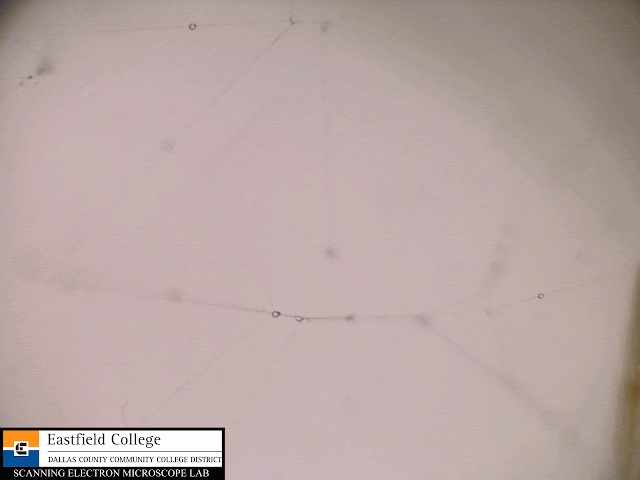
While in the collecting vial overnight, the spider spun a cobweb. 
This image shows the sticky droplets that help capture prey.

Well hello there!  A first portrait.
The spider has been preserved in 70% propanol with a little glycerol added

This is a ventral view (the under side of the spider).
You can see the two body sections, the cephalothorax or prosoma, and the abdomen or opisthosoma.
Also clearly visible are the 4 pairs of legs and an anterior pair of pedipalps.
Notice also that the spinnerets are not at the end of the abdomen, but are underneath the spider. This was very helpful in identifying the spider as belonging to family Theridiidae.


Pedipalps with claws visible.  You can also see the chelicera and fangs.


Side view showing downward facing spinnerets and narrow pedicel that connects the prosoma and opisthosoma.


Eye arrangement is important in identifying spiders.  This first look threw me off the trail since it looks like the spider only has 6 eyes.  It turns out, and it is obvious under the SEM, that the outer most eyes are paired - a dyad.
There are two rows of eyes, the anterior eye row and the posterior eye row.  In addition there are anterior median eyes (AME), anterior lateral eyes (ALE), posterior median eyes (PME), and posterior lateral eyes (PLE).  On this spider the the ALE and PLE form the dyad.

Spiders exchange gases via a highly folded book lung. 
I got lucky with the light and was able to photograph the book lung as indicated by the black arrow.


Spiders spin an egg sac to contain their eggs while they develop. 
I collected two egg sacs to image.  Notice the little spiderling outside of the egg sac (red arrow).

I didn't want this posting to be too long so it contains no scanning electron micrographs. I will post those images tomorrow, so please check back.

If you are interested in doing spider work let me recommend some books.
  • How to Know the Spiders by B.J. Kaston, 0697048993  This is the classic from 1978 and I would suggest beginning with this book since it is inexpensive.  Updates to the book are available online.
  • The Biology of Spiders by Rainer Foelix, 3rd Ed., 0199734828  Not an identification guide, but will tell you so everything you want to know about spiders.
  • Spiders of North America: An Identification Manual by Cushing and Ubick, 0977143902  This guide is Kaston on steriods.  Pricey, but well worth it for identifying spiders.
Eastfield College is dedicated to supporting STEM research at all levels.  If you would like to become involved with the Scanning Electron Microscopes at Eastfield please contact me.

Murry Gans
Scanning Electron Microscope Lab Coordinator
Eastfield College
Mesquite, TX
mgans@dcccd.edu
972-860-7267

Monday, August 13, 2012

Geology on a small scale


I am constantly searching for new and interesting things to image with the scanning electron microscope here at Eastfield College.  Last week I ran into one of the geology faculty members and asked for some samples I might use.

He told me an interesting story - while he and his students had been out collecting, they came across a formation of chalk that had some sort of burrow in it that had then been filled in with minerals that had crystallized.  He wasn't sure if  the burrow had been dug by some sort of worm or if it had been from a tree root.  He loaned me a couple of pieces for the SEM.

(Note:  You can click on any of the images to get a slide show of the images.)

 A photo of my "burrow" samples

Photo of the sample end on.  This is a bifurcation point - one branch leads off to the bottom right. 
On this image you can also see some of the chalk left on the sample.

For a closer look I imaged my samples on a dissecting scope.

Better light from the dissecting scope shows some great colors.
  
A side view of  one of the samples.  Very interesting cubical structures.
  
Another side view.  (The pinkish blob in the top left corner is my finger.)

I know absolutely nothing about geology so I had no idea what to expect.  I was unable to determine the origin of the burrow with the SEM, but I did discover some amazing structures. The images below were made by looking at the end of the bifurcated specimen.
   
[150x]
Lots of facets and layers
 
[454x]

[150x]
The first thing to catch my attention was the cubic box in the center of this image.


 [320x]


 [750x]

I discovered the smoother structures shown in the next two images.  What turned out to be even more interesting are the needle-like crystals and small spheres on the edge of the images. 

The SEM captures images by making a slow scan which allows me to see the image being formed line by line.  This is often how I discover new structures to investigate.

[1,000x]

 [2,700x]
This structure has a hole in it.  Notice the needle-like crystals to the left.


[3,200x]
A closer look at the needle-like crystals. 
Distance between marks on the scale above is 1 micro.

 [1,400x]
Note the pyramids and spherical structures.

[20,000x]
The square base of the pyramid is 1.7 micros on each side.
Distance between marks on the scale is 0.2 microns.

The images that follow are of the structures on the side of the specimen.  Most obvious are the cubic structures, but I was soon to discover more micro structures.


[50x]
While this image was forming, I noticed the hexagonal structure in the top left of the image.
 
[150x]


[745x]
Hexagonal faces and square faces.
On the right side you can see the chalk that was not cleaned away

[2,000x]
Chalk

[4,140x]
Chalk
 
[1,400x]
Plate-like structures underneath

[4,000x]

[1,400x]
More plates - notice the repeating "T" structures.

[4,000x]
Close up of a "T" structure

[1,500x]
Spherical structures
 These are some pretty remarkable structures - really makes me wish I knew some geology.  More important, images such as these could get your students interested in geology - especially if they make these "discoveries" themselves. 

Eastfield College operates two scanning electron microscopes that can be used by your students at no cost.  The mission of the SEM Lab is to encourage students go into the STEM fields and to consider coming to Eastfield to begin their undergraduate studies by giving them the opportunity to use our scanning electron microscopes .

The link below will introduce you to our portable SEM and show you how easy it is to use.

 
 I would love to talk to you about bring the TM1000 SEM to your school, having you come visit the Scanning Electron Microscope Lab at Eastfield, or collaborating with you and your students online.

Murry Gans
Scanning Electron Microscope Lab Coordinator
Eastfield College
Dallas County Community College District
Mesquite, TX
972-860-7267