
I have been working with the scanning electron microscope at Eastfield College for about four weeks and have finally made some progress on high magnification with high resolution.
Today I selected a flower from one of the planters on campus - a penstemon I believe.

I took one of these pretty little flowers back to the lab and used our Leica dissecting scope to make some images.


Now on to the SEM. (The image below is inverted so it is in the same orientation as the one above.) Right away you see that the resolution and depth of field with the SEM are pretty sweet!
But what about those black specks in the middle of the image? Pollen!

Part of a stamen. (190x)

Pollen grains. (1000x)

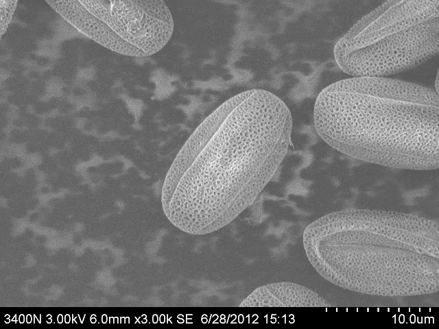
Pollen grains. (3000x)
Pollen grains. (5000x)

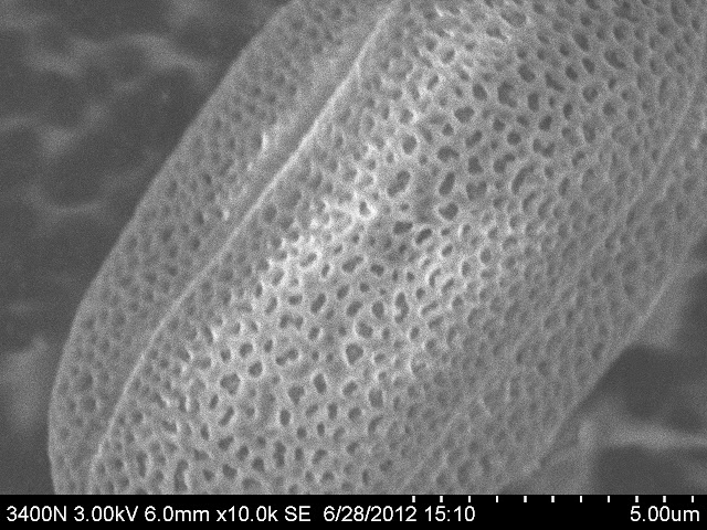
Pollen grains. (10,000x)
I took me several hours, but these are the best high resolution, high magnification images I have made with the SEM. It turns out to be a bit of an art, but I am learning.
A reminder that I am looking for science classes that would like to see and use the scanning electron microscopes at Eastfield. I am particularly interested in students in the Dallas / Fort Worth area who might be interested in attending Eastfield.
Spread the Word!
No comments:
Post a Comment