
When people find out that I am an electron microscopist they invariably asked if I have imaged bacteria, so I thought I would give it a try and see what I could accomplish.
In electron microscopy the degree of sample preparation is directly related to the quality of the images created. Even though we have outstanding scanning electron microscopes in the lab, we do not (yet) have a sputter coater or a critical point drier. The critical point drier would allow us to remove all of the water from specimens without changing their shapes and the sputter coater would add a very thin layer of gold and palladium to the outside of the specimen greatly increasing the amount of information the SEM can pick up.
In electron microscopy the degree of sample preparation is directly related to the quality of the images created. Even though we have outstanding scanning electron microscopes in the lab, we do not (yet) have a sputter coater or a critical point drier. The critical point drier would allow us to remove all of the water from specimens without changing their shapes and the sputter coater would add a very thin layer of gold and palladium to the outside of the specimen greatly increasing the amount of information the SEM can pick up.
These two machines are expensive and, while I hope to add them to the lab at some point, right now they simply aren't available to us. But that doesn't keep me from imaging - I just do the best I can with what I have.
Trisha, Eastfield's Biology Lab Coordinator, was kind enough to provide cultures of Micrococcus luteus and Bacillus subtilis for me to play with. She maintains bacterial cultures for our microbiology lab and had these on hand - they are also non-pathogenic.

I learned from Dr. B. Armbruster from Hitachi that using a transmission electron microscope grid as an easy way to mount bacteria for the SEM. The TEM grids are normally used to support the ultra-thin sections used in the TEM. I simply dunked the grid (dunked is a scientific term, right) into the water/bacteria mixture, let it dry, and then mounted it on the regular SEM stub.
 |
| The TEM grids are very small - you have to handle them with fine forceps. (Notice the torn grid at the bottom of the picture. I did that picking it up.) The large, ridged object on the left-hand side of the image above in the tip of my index finger. |
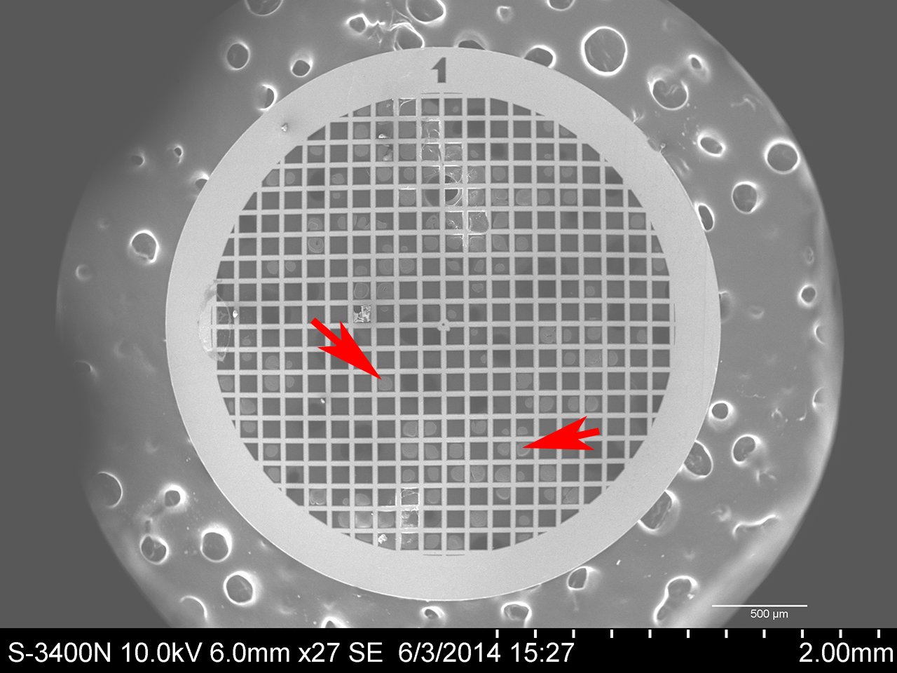 |
| Here is an image of the TEM grid in my scanning electron microscope magnified 27x. The arrows indicate areas where the bacteria have formed on the film between the square supports of the grid. Those spots contain lots of bacteria - way too many. I imaged in areas of the grid that had the fewest cells. |
Micrococcus luteus
Micrococcus luteus is a Gram-positive coccus that is found on human skin as well as in water, dust, and soil. Micrococcus is generally non-pathogenic, but can cause problems in people with compromised immune systems. (https://microbewiki.kenyon.edu/index.php/Micrococcus) This image was made by combining the images from two different detectors on the microscope, the secondary electron detector and the backscatter detector.
 |
| Micrococcus luteus [6,020 x]
This is one of the first images I took of M. luteus and I was very pleased with it. The white dots on the cells I believe to be charging caused by the electron microscope. These are prokaryotic cells and don't have membrane-bound nuclei.
|
Another pretty cool thing the SEM can do is mix signals from the two different detectors as different colors. I chose one detector to be red and the other to be blue. The result are some very lovely purplish bacteria.
 |
| Micrococcus luteus [10,000 x] |
 |
| Micrococcus luteus [37,000 x]
To get this image I maxed out the voltage on the SEM - 30,000 volts. Diplococcus is one form M. luteus can take (dipl- Greek diploos, double; cocc- Greek kokkos, a kernel or grain.) The "dots" on the surface of the cells are still there, but there was no charging. I have no idea what they are.
|
 |
| Micrococcus luteus [30,100 x] - Tetrad
Micrococcus luteus often occurs as a tetrad or group of four cells.
|
 |
| What the heck. Fun with Photoshop! |
Bacillus subtilis
Bacillus subtilis cells are rod-shaped, Gram-positive bacteria that are naturally found in soil and vegetation. (http://microbewiki.kenyon.edu/index.php/Bacillus_subtilis)
These bacteria gave off electrons at a higher rate and were much easier to image.
 |
| Bacillus subtilis [3,730 x] |
 |
| Bacillus subtilis [6,500 x]
B. subtilis is about 2 microns by 0.7 microns. This image was made with only the backscatter detector. Compare to the image below, which is at higher magnification, but of better quality because it is a composite of signals from two detectors.
|
 |
| Bacillus subtilis [11,000 x] Secondary electrons plus backscatter electrons. |
 |
| Bacillus subtilis [11,000 x] Same image as above, but with color mixing. |
 |
| Bacillus subtilis [27,500 x] |
One last note for any students who might be reading. Notice that scientific names - genus and species - are always italicized and that the genus name is always capitalized while the species name is never capitalized. After you use a scientific name for the first time in whatever you are writing, it is common and acceptable to shorten the genus to a single letter. Bacillus subtilis becomes B. subtilis. Do this and your professors will think you know what you are doing. Don't do it and they will not smile kindly upon you.
 All images in this blog are covered by a Creative Commons Attribution - Non-Commercial 4.0 International License. Feel free to used, copy, download, or modify the images anyway you like, just please give credit to Eastfield College, Mesquite, TX and don't sell them.
I welcome any comments on this blog.
Murry Gans
This is so awesome!! (I need to come up with better adjectives) I just took genetics and we talked in detail about prokaryotic organisms. Its cool to get to see them imaged! And I know they live on our skin and everywhere and outnumber us but its still so weird to see it magnified and so physical...if that makes sense!
ReplyDeleteI like the word "awesome"! Feel free to use it all you like.
ReplyDeleteI am delighted you like the blog so much and really appreciate that you take the time to comment.
Thanks for sharing. Learn a lot from your Blog.I really enjoyed reading it, you may be a great author.I must say you've done a wonderful job by sharing your article with us. Scanning Electron Microscopy
ReplyDeleteIn electron microscopy the degree of sample preparation is directly related to the quality of the images created. Even though we have ... eltmicroscope.blogspot.com
ReplyDelete