I am constantly searching for new and interesting things to image with the scanning electron microscope here at Eastfield College. Last week I ran into one of the geology faculty members and asked for some samples I might use.
He told me an interesting story - while he and his students had been out collecting, they came across a formation of chalk that had some sort of burrow in it that had then been filled in with minerals that had crystallized. He wasn't sure if the burrow had been dug by some sort of worm or if it had been from a tree root. He loaned me a couple of pieces for the SEM.
(Note: You can click on any of the images to get a slide show of the images.)
 |
| A photo of my "burrow" samples |
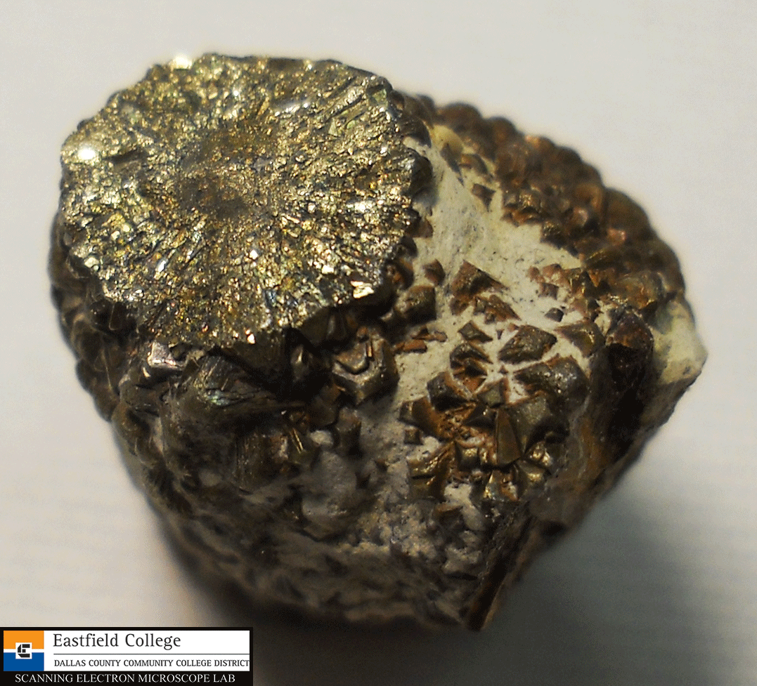 |
Photo of the sample end on. This is a bifurcation point - one branch leads off to the bottom right.
On this image you can also see some of the chalk left on the sample.
|
For a closer look I imaged my samples on a dissecting scope.
 |
| Better light from the dissecting scope shows some great colors. |
 |
| A side view of one of the samples. Very interesting cubical structures. |
 |
Another side view. (The pinkish blob in the top left corner is my finger.)
|
I know absolutely nothing about geology so I had no idea what to expect. I was unable to determine the origin of the burrow with the SEM, but I did discover some amazing structures. The images below were made by looking at the end of the bifurcated specimen.
 |
[150x]
Lots of facets and layers
|
 |
[454x]
|
 |
[150x]
The first thing to catch my attention was the cubic box in the center of this image.
|
 |
[320x]
|
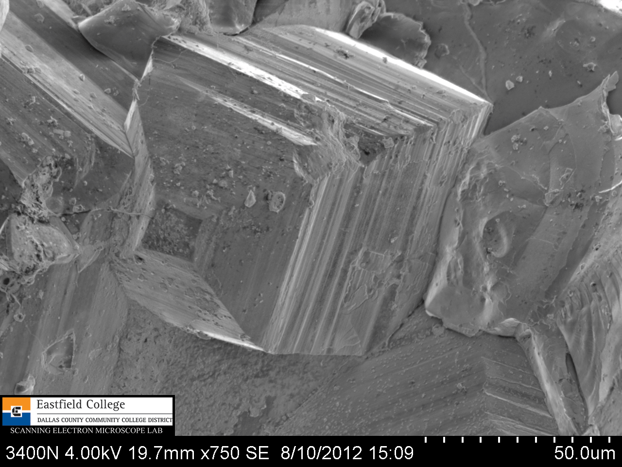 |
[750x]
|
The SEM captures images by making a slow scan which allows me to see the image being formed line by line. This is often how I discover new structures to investigate.
 |
[1,000x]
|
 |
[2,700x]
This structure has a hole in it. Notice the needle-like crystals to the left.
|
 |
[3,200x]
A closer look at the needle-like crystals.
Distance between marks on the scale above is 1 micro.
|
 |
[1,400x]
Note the pyramids and spherical structures.
|
 |
| [20,000x] The square base of the pyramid is 1.7 micros on each side. Distance between marks on the scale is 0.2 microns. |
The images that follow are of the structures on the side of the specimen. Most obvious are the cubic structures, but I was soon to discover more micro structures.
 |
[50x]
While this image was forming, I noticed the hexagonal structure in the top left of the image.
|
 |
| [150x] |
 |
| [745x] Hexagonal faces and square faces. On the right side you can see the chalk that was not cleaned away |
 |
| [2,000x] Chalk |
 |
| [4,140x] Chalk |
 |
| [1,400x] Plate-like structures underneath |
 |
| [4,000x] |
 |
| [1,400x] More plates - notice the repeating "T" structures. |
 |
| [4,000x] Close up of a "T" structure |
 |
| [1,500x] Spherical structures |
Eastfield College operates two scanning electron microscopes that can be used by your students at no cost. The mission of the SEM Lab is to encourage students go into the STEM fields and to consider coming to Eastfield to begin their undergraduate studies by giving them the opportunity to use our scanning electron microscopes .
The link below will introduce you to our portable SEM and show you how easy it is to use.
I would love to talk to you about bring the TM1000 SEM to your school, having you come visit the Scanning Electron Microscope Lab at Eastfield, or collaborating with you and your students online.
Murry Gans
Scanning Electron Microscope Lab Coordinator
Eastfield College
Dallas County Community College District
Mesquite, TX
972-860-7267
No comments:
Post a Comment