
Thursday, November 8, 2012

A Jumping Spider


An English professor here at Eastfield wanted her students to look at the world in a new way so she brought them to the SEM Lab to see how images are made.  She required each of her students to come to the lab and take at least three images that they would use for the basis of a story and video.  Pretty innovative and cool.

One of her students brought a jumping spider to the lab for imaging.  He spotted the spider on the trunk of a tree and tried to take its picture.  Interestingly, he soon found that this little spider was very good at staying on the opposite side of the tree.  Once the student got a closer look at the spider in the lab it was instantly obvious why this spider is so good at playing hide and seek.

Dissecting Scope Image
Full body view of jumping spider - yes, he is missing his second leg on the right side.

Dissecting Scope Image
What first caught our attention were the irridescent chelicera.  The student was quite amazed to see the large, forward facing eyes of the spider.

Dissecting Scope Image
Six of the spider's eight eyes are visible in this image.  The two large anterior median eyes, the two smaller anterior lateral eyes, and on either side of the head two posterior lateral eyes


The next two images are my favorites.  I tipped the spider face up to get a front-on portrait.  If you are a prey insect, this is the last thing you will ever see.

Dissecting Scope Image

Dissecting Scope Image

The following images were made with the scanning electron microscope.  As always, they are not in color because they are not made with light.  Not as pretty, but greater depth of field and resolution.
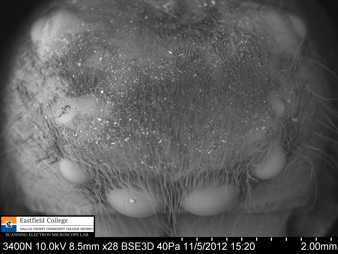
SEM Image [28x]
All eight eyes are visible in this image.  They form a big "U".

 SEM Image [35x]
Anterior eyes

SEM Image [21x]
OK, so I have a thing for spider faces.

 SEM Image [42x]
Well hello there!

SEM Image [52x]
Salticids, jumping spiders, have excellent vision and can see for several centimeters.  One of my spider books says that their behavior is very similar to that of cats.

This was a student's specimen, so I didn't do a detailed study with the SEM.  I was looking for unusual things that he might find interesting. (I also wanted to show you that I could take images that were not spider faces.)
 SEM Image [94x]
Jumping spiders are HAIRY!!  I love everything about this image of the leg of the spider.
Who would have imagined?

 SEM Image [320x]
This is an image of the anterior surface of the abdomen of the spider.  Honestly, I have no idea what the function of the scale-like hairs might be. 

I have always thought spiders to be among the most bizarre and interesting of animals.  The more I get to see them "up close and personal", the more intrigued I have become.

Eastfield College is dedicated to supporting STEM research by students at all levels.  I welcome your inquiries and comments.

Murry Gans
Scanning Electron Microscope Lab Coordinator
Eastfield College
Mesquite, TX
972-860-7267


Wednesday, October 17, 2012

A Kidney Stone - OUCH!!


I have done my best to make sure that everyone knows that Eastfield College has a Scanning Electron Microscope that students and faculty can use.  In fact, if you read this blog at all, you know that I say that in every post. (Oh, look. I just did it again!) As a result several students show up to use the SEMs each day and I am beginning to take our portable SEM to area schools. 

One thing that I really didn't expect is people bringing me things to image, and today I was handed my most interesting specimen - and I do mean specimen.  A kidney stone that one of my colleagues passed yesterday.  (I will not tell you who - he or she can tell you if they want to.)

I am told that passing a kidney stone is an extremely painful experience.  The images below really show why that is true.

So brace yourself for an up-close and personal look at a kidney stone.

 Dissecting Scope Image
This is not the entire stone - a small piece broke off

Dissecting Scope Image

SEM Image[20x]

SEM [249x]
Take about death by a 1000 cuts, this view down one edge really shows the angular crystals of calcium and uric acid.

SEM [850x]

SEM [500x]

SEM [208x]
Top view showing not only angular crystals, but also round nodules

SEM [750x]

SEM [250x]

SEM [400x]

SEM [800x]

SEM [2,000x]

SEM [65x]
Interestingly, part of the kidney stone was not made of sharp and nodular crystals.  There was a more solid portion.
SEM [200x]

SEM [561x]
This image, and the one below, show some of the cost of a kidney stone.  The dark patches are tissue from the donor.
SEM [1,600x]

Definitely one of the most unusual and scary things I have imaged.  I don't know about you, but I am going to drink more water!!

See you in the lab.


Friday, October 12, 2012

My Favorite Harbinger of Fall

The Late Blooming Aster

(Symphotricum patens - I think)


I love warm weather, but Texas summers are often pretty brutal. I am always delighted when the break in the weather occurs in the fall and our temperatures drop from highs in the 100's to highs in the 80's - often over night. 


When this little aster shows up in my yard and I know that fall is near.  Now that the temperatures have finally changed for the better, these asters are everywhere. 

With flower heads only about 5/8ths of an inch across, these little guys are often over looked, but if you take the time to really look at them, you are in for a treat.  The petals are white to lavender.

 Dissecting Scope Image
This flower head is in a small jar filled with water to keep it turgid. 

Dissecting Scope Image
This is not a simple flower, but a composite flower - made up of several individual flowers.  The flowers in the middle are called disk flowers and have no petals.  The flowers on the outside are called ray flowers and have only a single petal.  If you count the petals you will know how many ray flowers there are.

Dissecting Scope Image
A close up of the disk of this composite flower.  Notice on the image above that 8 of the disk flowers have yet to open.  The little dots in the image are pollen grains.

Disk Flowers - SEM [20x] 
Even though this is only a 20x magnification, the SEM provides excellent depth of field which allows almost the entire flower disk to be in focus.  The flowers in the middle are not open. The flowers farther out have opened and the anthers are protruding.  These will also open up and a stigma will emerge from the center.

 Outer covering of anthers (pollen obvious) - SEM  [120x]

 Anthers - SEM [25x]
On the bottom left of the image you can see a stigma from a ray flower.

 Dissecting Scope Image
Pollen on anther

Composite flowers can be difficult to figure out, so I took the flower head apart to look at the disk and ray flowers.

 Dissecting Scope Image

 Dissecting Scope Image
This is a ray flower with its single petal.  Seed dispersal in this aster is just like a dandelion - once the seed forms in the ovule it will be dispersed by wind.  The pappi in the image will become the parachute that carries the seed.

Dissecting Scope Image

A close up of the stigma of a ray flower.  I am very pleased with this image - you can clearly see the pollen grains on the stigma

But what about those stamen?  They really don't look like the stamen on simple flowers.  In the images below I have used a needle to dissect a disk flower to find the stamen.

 Dissecting Scope Image
Even on this low magnification image you can see that the anthers on the inside of the flower.

 Dissecting Scope Image
An excellent image showing pollen lined up on an anther.

 Dissecting Scope Image
With the disk flower fully open, the stamen are finally revealed.  The anthers are covered in pollen and supported by filaments.

 Tip of unopened stamen - SEM [55x]
The pollen is essentially oozing out of the stamen

Close up of stamen with pollen - SEM [185x]

 Anther with lots of pollen - SEM [62x]

 Filaments that support anthers, Pappi - SEM [71x]

The final two images are of pollen.

 Pollen - SEM [750x]

 Pollen - SEM [1,000x]
Pollen about 25 microns in length - small!

It was a lot of fun to study the composition of this aster using the tools available in the Scanning Electron Microscope Lab here at Eastfield College.  It is amazing what you discover when you are able to see the world in such tiny detail.

The SEM Lab at Eastfield College is open to students and faculty everywhere.  We have two scanning electron microscopes.  Our smaller scope is so simple to use that I can have you capturing images within 15 minutes.  The larger scope provides more flexibility and control, and I am here to help you use it. 

If you are interested in seeing or using our scopes, please contact the lab.

Murry Gans
Scanning Electron Microscope Lab Coordinator
Eastfield College
Mesquite, TX