Wednesday, August 1, 2012

A Lone Drone (Bee)


A lady who works across the hall from the SEM lab is an amateur bee keeper. I asked her to keep an eye open for me and bring in any bees that she finds dead - on Monday she brought me a drone.  That in itself is pretty cool since all of the bees you see around flowers are workers.

Of course, this little drone bee went right into the scanning electron microscope for a pretty interesting afternoon of imaging.

 Talk about a head shot!! 
Eyes, antennae, mouth and first pair of legs visible.
[20x]

Notice that this hairy guy even has hair on his eyes! 
[36x]
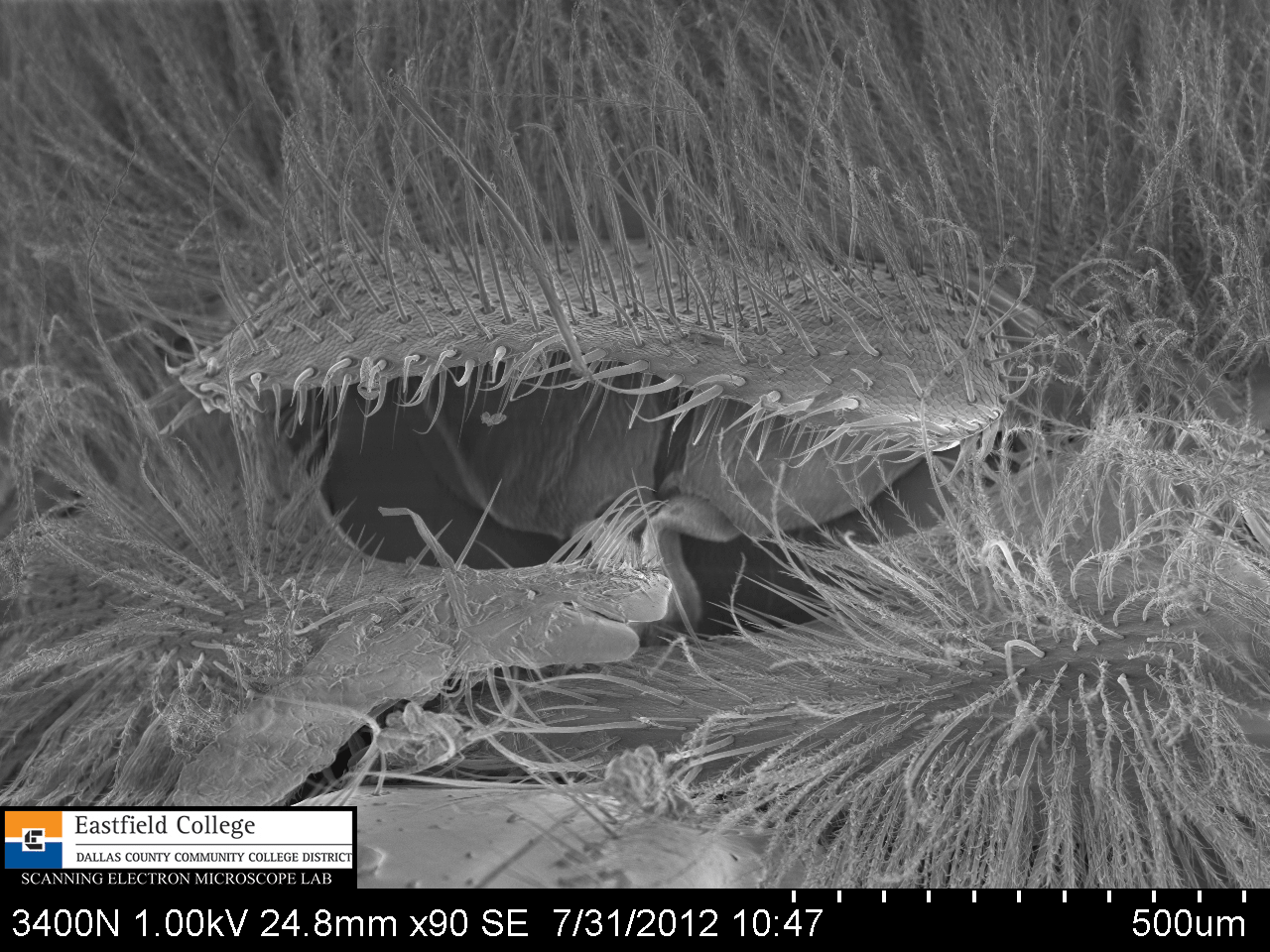
A closer look at the structure in the middle of the head - the labrum or upper lip 
[90x]

The mouthparts of the bee
The glossa, or tongue, extends downward
[55x]

 The right maxilla of the bee
[141x]

 Appendage on maxilla
[160x]


Maxillar appendage  
There appear to be sensory structures present - a focus for further imaging. (No pun intended)
[274x]

The business end of the glossa, or tongue 
[160x]

The most distal 5 segments of the bee's antenna
[80x]

 Terminal segment of an antenna
[210x]


Antenna segment
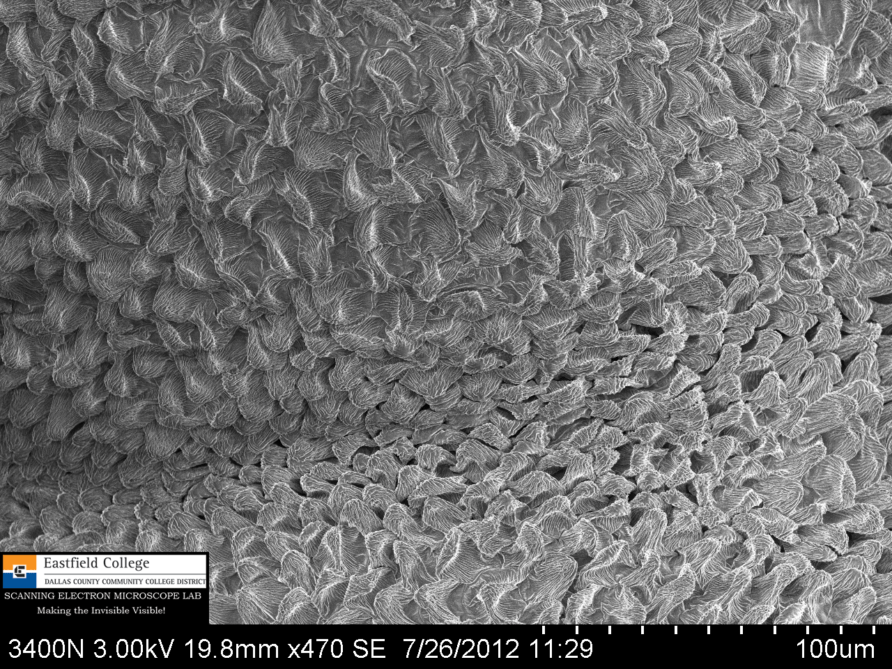
[470x]


 I noticed that many segments of the antenna are pourous.
This is really obvious along the right hand side of this antenna.
Note in this image the interesting patch of black spots in the upper left of the image
[745x]

 The black patch actually contains what appear to be 5 different types of receptors
More questions to be answered.
[1,800x]

 Now at look at that hairy, compound eye
[39x]

Ommatidia and hairs
[143x]

I have now been a scanning electron microscopist for about 8 weeks.  I remain amazed (yes, I know I say that a lot, but it is true) at what the SEM can show me.  Most important, the observations I have been making have sent me to the internet and library find out more about what I am seeing. 

Having access to high quality tools can go along way to stimulating a life long interest in science - not just biology, which is obviously my preference, but also physics, chemistry, geology, and forensics.

The electron microscopes at Eastfield are available to teachers and students at all levels.  If you would like to do some imaging please contact me.

Murry Gans
Scanning Electron Microscope Lab Coordinator
Eastfield College
972-860-7267


Friday, July 27, 2012

A Flesh Eating Predator (Less than 3 mm long)


In my previous blog post I showed images of Lantana montevidensis.  When I brought the first sprig of Lantana back to the lab a small spider came along for the ride. Because so many different kinds of insects live on and in the Lantana, it makes sense that it would be a good a niche for a tiny predator.

This spider is less than 3mm from the most anterior part of its cephalothorax to the posterior end of its abdomen.

This is not a very detailed study because this is the first spider I have tried to image with the scanning electron microscope.  I had a problem with the build up of charge on the spider (which distorts the image). I will have to do some research and experimentation to figure out how to keep this from happening. 

Never the less, the following images should give you a feel for the marvelous strangeness of spiders - some of my most favorite creatures on the planet. 

Spiders this size can be frustrating to study - I have tended to avoid them. 
Now, thanks to my access to an SEM that will change. [22x]

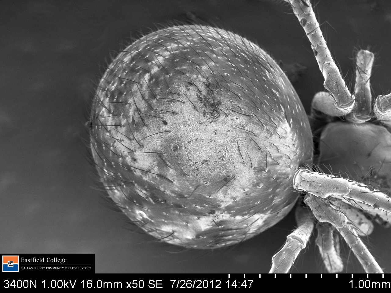
A closer look at the abdomen [50x]
You may notice the absence of spinnerets on the posterior end.  On this species they are on the ventral surface of the abdomen.

 At 177x ridges on the abdomen become visible.


Dorsal Abdomen [384x]

 A look at the pedicel that connects the cephalothorax (right) to the abdomen (left) [255x]


 Cephalothorax and eyes [90x]

 Cephalothorax and eyes [160x]
Ridges become apparent


Talk about a head shot!  [471x]
I wonder where they got the ideas for Spider Man's costume?

 Femur of the hind most leg on the right side of spider [350x]

Patella at top, Tibia below [200x]
Look carefully and you can see three distinct type of hairs. 

Close up of hair types on tibia [903x]
Affect of charging is obvious - I am going to work on this.

Many years ago I had the opportunity to develop and teach an arachnology course to high school students.  I will be honest, studying spiders is difficult, but we made some progress and my students and I really enjoyed it.  When I changed schools, unfortunately, there was no interest in the class, so I haven't done a lot of spider work for some time.  Chancing upon this little spider has definitely rekindled my interest.

If you would like a chance to see and use a scanning electron microscope to image spiders or anything else, please don't hesitate to contact me.  Eastfield college is dedicated to supporting work in the STEM fields at all levels - for both faculty and students.  If you are not located near Eastfield College, we can still collaborate online.  I Skype.  Hope to hear from you soon.

Murry Gans
Scanning Electron Microscope Lab Coordinator
Eastfield College
972-860-7267


It All Begins Here!


Lantana montevidensis


On my way the administration building I came across what my wife and I call butterfly weed.  My online research reveals it to be Lantana montevidensis (at least that is what I think it is). 

http://eol.org/pages/579777/overview

It turns out that it is not a butterfly weed at all - that will teach me to use a common name - especially one I make up.

I reach down and cut off a sprig with my pocket knife and head back to the lab, then return with my camera to get a few pictures


It may not be a butterfly weed, but butterflies sure like it. 
This swallowtail came for visit while I was taking pictures.


_______________________________________________
Next it is on to our dissecting scope for a closer look.

Nice long tubular flowers - made for butterflies, and butterflies made for it!

 Butterflies aren't the only insects that like this plant.  On this close up you can see a small insect in the picture.  I was lucky to catch him - the capture rate on the camera is pretty slow.  By the time I could take a picture he would scurry away. (The colors you see on the insect here are because of chromatic abberation - very pretty though.)

Finally got him.  True colors this time.  I don't know his genus and species, but he is definitely a hemipteran or true bug.

 Flower head straight on.  Notice the small hairs just inside the tube. 
You will see these again on the scanning electron micrographs.

 Yet another resident on this flower living inside the tube of the flower.


 Here is a leaf from plant - just for the record.

_______________________________________________
On to the SEM.  I didn't collect and image the insects.  I actually work backwards - SEM first, then camera and dissecting scope.  I didnt' even know the insects were there until these images were made.  I wouldn't have collected the butterfly anyway - it is hard to kill something that beautiful.

 For this image I split the flower open. 
You can see the layer of hairs just inside of the tube and farther down are 4 stamen. [13x]

 In this image you can see the stamen, another field of hairs, and then the stigma.  [25x]

 Stamen [55x]

A close look at the cells that make up the tube of the flower. 
This is a close up of the section seen on the right hand side of the image above. 
At the bottom is one of the "hairs" inside the tube. [691x]

Cells of the stamen.  Where is the pollen?  [150x]

 Make up of a petal - the part in focus is the outside of the petal folded back on itself [101x]

Close up of cells that make up the petals.  [470x]

 Upper surface of leaf [60x]

Closer look at upper surface of a leaf [250x]. 
I simply cannot believe how alien this appears.

 Bottom leaf surface. 
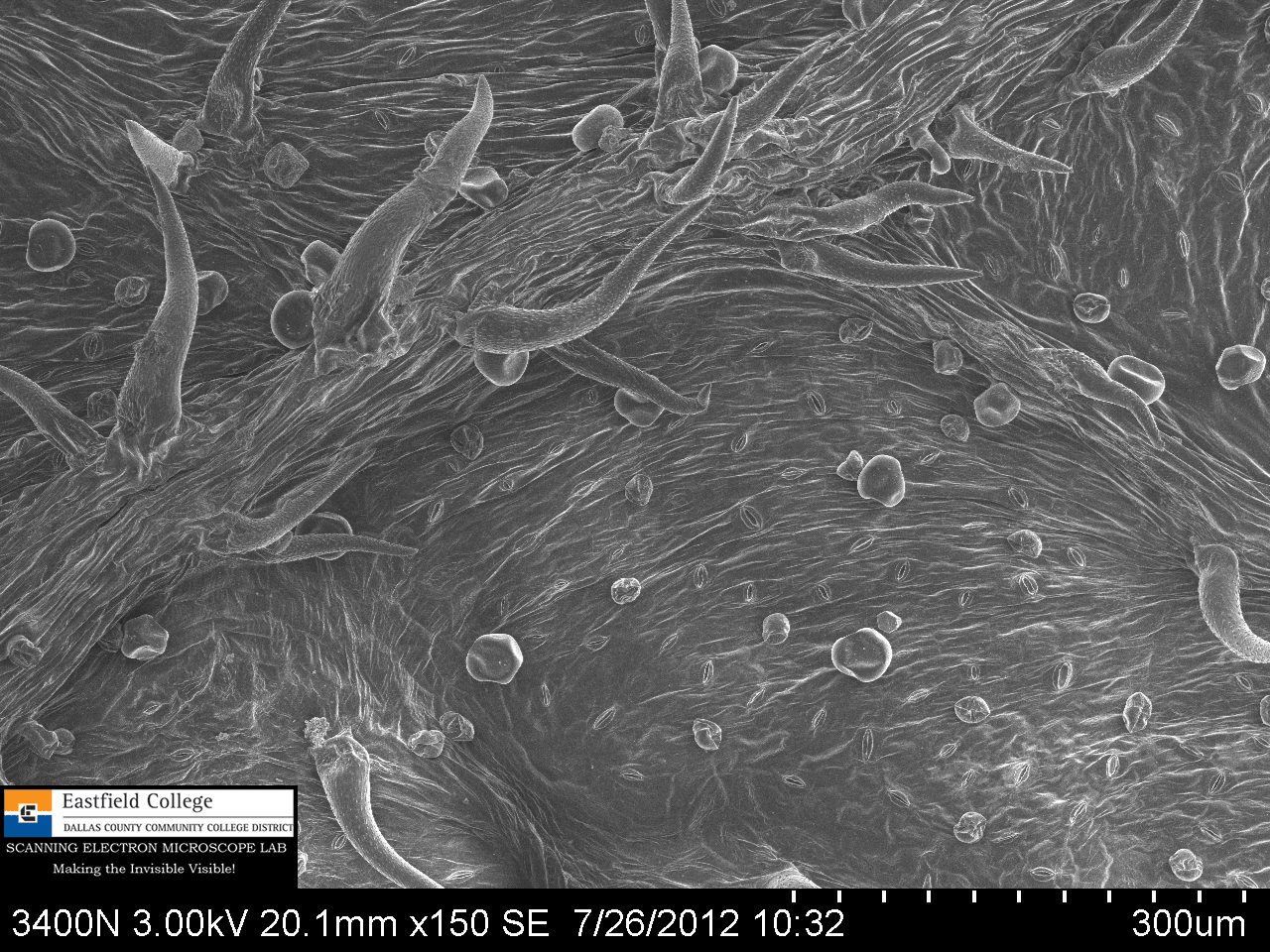
The tree-like structure running left to right is the midrib.  Stomata are visible.
The globular structures must be glands that contain aromatic liquids. [150x]

 Bottom leaf surface [250x]


Stomata on bottom of leaf [800x]
Distance between marks on scale = 5 microns

I welcome you comments on this posting.

If you are interested in using the SEMs at Eastfield or would like for me to bring the portable SEM to your school, please contact me. 
Eastfield is dedicated to the support of research in the STEM fields by students and faculty at all levels.

Murry Gans
Scanning Electron Microscope Lab Coordinator
Eastfield College
972-860-7267


It All Begins Here!