Tuesday, July 24, 2012

Copper Wire - Cooler than it sounds (at 5,000x)


An undesirable phenomenon called charging can occur when using a scanning electron microscope.  The electrons from the electron gun need a conductive pathway.  Nonconductive specimens will accumulate a charge - they become very bright on the screen.  The worst part is that at some point they will discharge which can distort the image.

There are lots of ways to deal with this, like reducing the gun voltage, but I wanted to try my hand at using some high voltages.  My solution - use copper wire. 

I found an old telephone line, opened it up and stripped the insulation off one of the small wires inside.  (I used a pocket knife - not such a good idea as you will see.)

Here are my lovely copper wires at 37x  (Gun voltage = 15kv)


A closer look at 1,000x


At 5,000x the smooth wires are looking a little rough.
Notice the striations that show where the wire was drawn through the die.

____________________________________________________________
All of the images above were made using the secondary electron detector, thus the "SE" on the bottom of the picture.  SE images are made in a high vacuum.  The problem is, I purposely left some the insulation on the wire.  Talk about charging!!  Another solution to the charging problem is to use the back scattered electron detector.  This detector can work in a partial vacuum.  That little bit of air can solve the charging problem.  Notice on the images below the "BSE" as well as the atmosphere in the scope.  In the next image shows an atmosphere of 6 Pascals.  (Standard atmospheric pressure is 101,325 Pa.)
____________________________________________________________

 Why you shouldn't strip wires with a pocket knife (42x)

 (110x)


Copper is soft.  Looks like I have effectively reduced the diameter of the wire by half. (400x)


 Back to SE mode at a lower voltage.  The surface of the wire shows what appears to be crystallization. (1,600x) I am going to have to go back to the lab and take another look. 

Have something you would like to see up close and personal?  Give me a call.

Murry Gans
Scanning Electron Microscope Lab Coordinator
Eastfield College
972-860-7267


Sunday, July 22, 2012

Popcorn Revealed


The scanning electron microscopes at Eastfield College are available for use by any student or faculty member free of charge.  In an effort to advertise this fact, once a week I take our small scanning electron microscope to the "Pit" so students, faculty, and staff can see it in action.

The Student Life organization at Eastfield is kind enough to come each week and give away free popcorn while I am there.  (It is dang good popcorn, by the way!)

Last week a young man, I am guessing Middle School aged, was eating in the Pit with his dad and came over to see what I was doing.  We dediced to put a piece of popcorn into the scope and see what it looked like.  Below are the images we made.

 Image 1:  Outer side of popcorn [30x]

Image 2:  Outer side of popcorn [250x]


Image 3:  Outer side of popcorn [1,200x]


 Image 4:  Inner side popcorn kernal [30x]


Image 5:  It is salted popcorn!! [250x]

It is pretty obvious that nature loves hexagons.  What first struck me was how much these exploding hexagons of starch look like the cells in a bee hive.  Bees build in hexgons because they can be tesselated - placed together with no wasted spaces - and because it allows them to build the largest volume for strorage with the least amount of material. 

I continue to be astonished by what the scanning electron microscope is able to show me.  Call and come by Eastfield and see what you can discover. 

Murry Gans
Scanning Electron Microscope Lab Coordinator
Eastfied College
972-860-7267

It all begins here!


Tuesday, July 10, 2012

A close look at a true bug


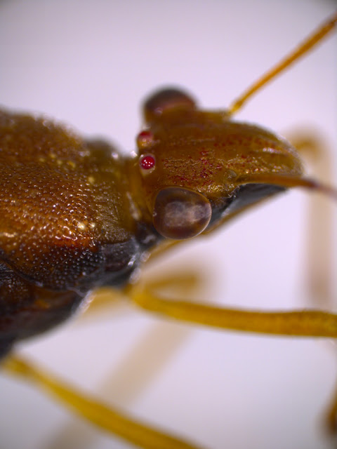
Back outside looking for insects, I came across this hemipteran, or true bug, on a Vitex tree on campus.  Hemiptera means "half wing" - even though hemipterans have two pairs of wings, when they are folded together on top of the body, the are half covered - a pretty easy way to identify the order.

By the way, this is the only order of insects that can be accurately be called "bugs".


In the picture below the compound eyes are obvious.  This insect also has a pair of simple eyes or ocelli - interestingly, on this bug they happen to be red.


This bug is eats the sap of plants using its piercing mouth parts.  If you look carefully at the image below you can see the long, piercing stylets tucked in along the midline of the body.


Now on to the scanning electron microscope. 
Below a look at the compound eye (95x).


A closer look.  (901x)  Each unit of the compound eye is called an ommatidium. For size refer to the scale at the bottom of the image.  On this image the distance between the white marks is 5 microns.


One of the simple eyes (174x). 
Scanning electron micrographs do not show colors - unfortunate in this case.


The piercing stylets of the mouth.  OUCH!


The business end of the mouthpart  (60x). 
This image also shows the connection of the second pair of legs to the thorax of the insect.


It turns out I have an obsession with the weird world of tarsal claws on insects. 
The next three images are of increasing magnifications of the tarsal claws on the first pair of legs. 

(200x)


A close up of the "pad" on the far right of the image above.

(850x)


These tubelike structures are just visible in the first tarsal claw image. 

(1000x)


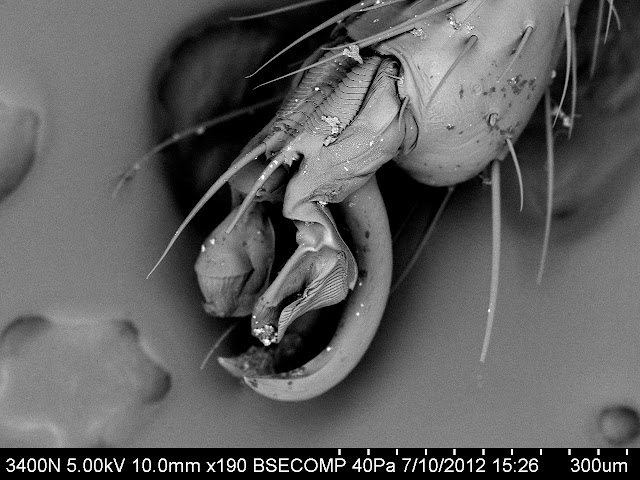
This tarsal claw belongs to one of the hindmost legs.
(95x)


A closer look at 190x. 
Notice the series of plates at the top of the claw and the two padlike structures - each is shown at high magnification in the images to follow.


Plates (500x)
[Talk about stranger than science fiction.]


Right side pad (700x)


Left pad (700x)


Awesomely strange, and lots of fun to discover.  If you are a student or teacher and would like to get involved with scanning electron microscopy the Scanning Electron Microscope Lab at Eastfield College is ready to collaborate.

Murry Gans
Scanning Electron Microscope Lab Coordinator
Eastfield College
Mesquite, TX
972-860-7267


Monday, July 9, 2012

An unexpected bonus - a thrip larva


In an effort to improve my skills with the electron microscope, I have begun to make detailed studies of some of the organisms on the Eastfield campus.  I picked the trees shown below because I could reach the leaves - most the of the established trees on campus have been trimmed too high for me.


I began with a dissecting scope and discovered some passengers on the  underside of the leaf that were too small for me to see with the naked eye (at least my naked eye).


I love those purple eyes!!


The thrip was interesting (of course at this point I had no idea what it was), but I was also intrigued with the leaves.  Notice on the picture below what appears to be some beads of fluid.  The plant was definitely aromatic giving off a strong but not unpleasant odor when I touched the leaves.


So now on to the scanning electron microscope.  First the upper surface of the leaf.

I am continually astonished at the strange worlds I find with the electron microscope - worlds that were there all along, in plain sight, yet still invisible to us.


It is just a guess, but I suspect that the four-part structure on the upper surface of the leaf shown below produces those little drops of liquid on the lower leaf surface.


Now for a look at the underside of the leaf.


The image below shows a vein on the leaf.  Who knew?


But what about that thrip??


The thrip's mouth parts point straight down.  Unfortunately, they are hidden by its right front leg. 


The bitter end.


In the picture below you can see a close up of the eye.  The large structures on the far right are antennae.


Now for the really cool part
I noticed a small structure between the first and second pair of legs on this little fellow.  The image below is magnified 400x - high power for a light microscope.


How about at 2,000x?  See below.


Now for 6,000x - six times oil immersion on a light microscope.


What the heck???  I will have to find out.

If you are near Eastfield College in Mesquite, TX, you and your students are invited to come and use the electron microscopes.  If you can't make it in person, I would be happy to work with you and your classes via Skype. Eastfields SEMs are also available for faculty and student research. 

Contact Murry Gans
972-860-7467

It all begins here!