Monday, December 8, 2014

Don't Fear the Flying Scorpions


In late October I made a quick trip to the Trinity River Audubon Center and came across a wonderful insect called the scorpionfly.  I was very pressed for time so only managed to capture a single specimen, but just a few days later Professor Jeff Hughes walked into my lab with another one.

The male scorpionfly.  Weird and scary looking, but totally harmless to humans.  They don't bite or sting.
Coming upon a scorpionfly for the very first time can be a bit of a surprise.  The elongated beak of the male is pretty weird looking, not to mention what appears to be a scorpion-like stinger.  But never fear - they are harmless.  In general, scorpion flies are scavengers that eat dead insects.  And the stinger is actually the male reproductive organ. (Which in a way makes it even more scary.) Scorpionflies belong to the order Mecoptera.


One reason I really like scorpionflies is that they are instantly identifiable, mainly because they remind me of my favorite part of Mad Magazine - the Spy vs Spy cartoons that were drawn in the margins of the magazine's pages.


The male genital structure of the Mecopteran.


Though called scorpionflies because they look like flying scorpions, but the scorpionfly is neither a scorpion nor a fly.  Flies have two wings while this insect has four, and scorpions are arachnids with 8 legs, not 6 as seen in insects.  
Scorpion flies are harmless to humans.  Most species either prey on small insects or eat the bodies of dead insects.  The long snout allows them to feed in narrow places and on small prey.

Mecoptera is a very old order of insects, dating back to the Permian period 299 to 251 million years ago. During the Permian, scorpion flies were the most abundant and diverse insects that went through complete metamorphosis and are probably ancestral to today's dipterans (flies) and lepidopterans (moths and butterflies).

This means they survived the Permian extinction - the largest mass extinction in the history of earth which allowed the rise of the dinosaurs -and then survived the asteroid impact 65 million years ago that finished off the dinosaurs. 


Here is a head-on view of the male scorpion fly as seen in the electron microscope.  You can clearly see the small aluminum wire I used for a mount.  The insect is attached to the wire with Elmer's Wood Glue.  It sticks to the chitinous exoskeleton well. SEM image. [10 x]

Now for some scorpionfly sex.

The male attracts the female by releasing pheromones from glands within the enlarged genital segment.  Before he does this he prepares a nuptual gift of a dead insect. When the female arrives she begins feeding and he grabs on to the edge of her forewing and while she eats he inseminates her.  (If only it were that easy for humans.)

This is the tip of the male genital structure.  Though I could find nothing about it, the function of the sensory hairs is obvious and I wouldn't be surprised to learn that sperm is inserted via the numerous holes.  SEM image. [320 x] 

I have known about scorpionflies for many years and have always enjoyed finding them, but recently the Eastfield library added a book called Planet of the Bugs by Scott Shaw on insect evolution that really opened my eyes to just how cool scorpionflies are.  If scorpionfly ancestors hadn't survived a couple of mass extinctions, we might not have flies and butterflies around today.  It is very cool to realize that these insects were buzzing around long before the dinosaurs and are still here.

My thanks to Michael Haas, a Ph.D. student at the  Department of Entomology at the State Museum of Natural History Stuttgart, Germany for pointing out a major error on the previous version of this blog posting.  I had identified a female Ichneumonid wasp, probably the genus Compsocryptus, as a female scorpionfly because of similar wing coloration.  I have removed those images from this posting to avoid confusion.

Science is based on the open exchange of ideas and information, and the internet provides a global reach.  Thanks for taking the time to let me know of my error, Michael.  I learned something new today.

Murry Gans
mgans@dcccd.edu
Eastfield College
Mesquite, TX

References:
Carde, R. T., & Resh, V. H. (2009). Encyclopedia of insects: Previous ed.: 2002. 
Amsterdam: Elsevier?Academic Press.

Kattes, D.H. (2009). Insects of Texas: A practical guide. College Station, TX: Texas A&M.

Shaw, S. R. (2014). Planet of the bugs: Evolution and the rise of insects. Chicago, IL:
University of Chicago.

Smith, D. (2011, June 30). The permian period. Retrieved December 8, 2014, from 
http://www.ucmp.berkeley.edu/permian/permian/php

Triplehorn, C. A., Johnson, N. F., & Borror, D. J. (2005). Borror and DeLong's introduction 
to the study of insects. Belmont, CA: Thompson Brooks/Cole. 


All images are covered by a Creative Commons License.  They may be downloaded, used, or modified with attribution to Eastfield College, Mesquite, TX, but may not be sold.

Tuesday, July 1, 2014

Sex in the weeds: When a young beetle's fancy turns to love.


My current focus in the lab is with Lixus scrobicollis, the ragweed weevil.  As I was walking up to my study site at the Trinity River Audubon Center in early June I saw what at first glance appeared to be bees hovering over the tops of the ragweed plants.  

This made absolutely no sense because the ragweed plants hadn't flowered and are wind pollinated - their flowers, when they make them, don't have petals.  

A closer look revealed not bees, but some small, orange beetles.  I watched the beetles land on the ragweed, but they weren't really showing any interest in it - they were just sitting there.  As I walked around more I discovered the beetles swarming on another plant called Illinois Bundle Flower (Desmanthus illinoensis) growing alongside the ragweed.  The females were happily feeding on the flowers and flower buds of the Bundle Flower, but the males were intensely interested in mating with the females.  The females seemed to be paying absolutely no attention the activity of the males.

I was so fascinated watching the beetles swarm, that, like a rookie, I didn't notice I was standing in a fire ant mound.  They quickly let me know.  After a few minutes of hopping around and trying to rid myself of fire ants, I moved to a better location and took some images.

Here is a short video I took of the action.


I have to  admit that I struggled with trying to identify the beetle for several days, but I am lucky because I know people who can help.  Dana See, a Lab Assistant in our biology labs is an outstanding naturalist and she came up with a classification, Anomoea flavokansiensis, in less than an hour.  

The scientific name is much longer than the beetle itself!

I asked her how she did it and she told me she did a Google search.  She picked much better search terms than I did so she wins.  I feel a little better since the genus Anomoea isn't in any of my entomology books.

Here are some images I took in the field.

In this image Anomoea is on a Saw-Leaf Daisy (Prionopsis ciliata).  This plant is also near the Illinois Bundle Flower and is just a resting place.


Anomoea resting on a ragweed leaf.  


In this image you can see a mating pair.  The female is happily munching away on the flower while the male copulates.

A look at the bipinnately compound leaves of the Bundle Flower.  The plants in the background are ragweed.

Mating pair on the left.  Other beetles under the leaves.

There are a lot of beetles in this shot - at least 4 mating pairs.


One of the best field pictures I have ever taken.  Notice the male's grip on the female with his tarsal claws.  

As you might have guessed, this beetle invasion may not be to the total benefit of the plant. Insects play a major role in plant pollination and there may be some pollination occurring here, but eating the flower reduces the ability of the plant to produce seeds.  
While I was observing the beetles, a group of young students came by my study site.  They were competing to see how many different species they could find so I showed them the beetles.  They quickly told me they had already seen them and wanted to know if I had anything else to offer. (Note to self - When talking to young people, don't point out that "the beetles are making more beetles" unless you are ready to explain how.  I wasn't and didn't!)

I found it interesting that these beetles were not on every Bundle Flower plant I saw that day. Why this particular plant and not others?  Location near water, maturity of the flower buds, detection of a chemical signal?  The female beetles are obviously interested in feeding on the flowers, but were both males and females attracted to the plant, or were the males attracted to the feeding females? Do females release a pheromone when they find a good source of food? If so, what is that pheromone?

Interesting questions, but for now my pursuit of ragweed weevils continues. 

This work is covered by a Creative Commons Attribution 4.0 International License. Images may be used or modified with attribution to Eastfield College, Mesquite, TX.  They may not be used commercially.

I hope you enjoyed seeing these little beetle making more beetles!  I saved some specimens for the scanning electron microscope and look forward to sharing those images with you.

I welcome any comments you may have.

Murry Gans
Microscopy Laboratory Coordinator
Eastfield College
mgans@dcccd.edu

Thursday, June 5, 2014

Imaging Bacteria in the Scanning Electron Microscope


When people find out that I am an electron microscopist they invariably asked if I have imaged bacteria, so I thought I would give it a try and see what I could accomplish.

In electron microscopy the degree of sample preparation is directly related to the quality of the images created.  Even though we have outstanding scanning electron microscopes in the lab, we do not (yet) have a sputter coater or a critical point drier.  The critical point drier would allow us to remove all of the water from specimens without changing their shapes and the sputter coater would add a very thin layer of gold and palladium to the outside of the specimen greatly increasing the amount of information the SEM can pick up.

These two machines are expensive and, while I hope to add them to the lab at some point, right now they simply aren't available to us.  But that doesn't keep me from imaging - I just do the best I can with what I have.

Trisha, Eastfield's Biology Lab Coordinator, was kind enough to provide cultures of Micrococcus luteus and Bacillus subtilis for me to play with.  She maintains bacterial cultures for our microbiology lab and had these on hand - they are also non-pathogenic.


I prepared the bacteria for the electron microscope by putting a small droplet of distilled water on a microscope slide and then, using an inoculating needle, transferred a minute amount bacteria to the water and mixed it up.  Just like in light microscopy, using too much bacteria is a bad idea - they all clump together making to hard to determine morphology.

I learned from Dr. B. Armbruster from Hitachi that using a transmission electron microscope grid as an easy way to mount bacteria for the SEM.  The TEM grids are normally used to support the ultra-thin sections used in the TEM.  I simply dunked the grid (dunked is a scientific term, right) into the water/bacteria mixture, let it dry, and then mounted it on the regular SEM stub.

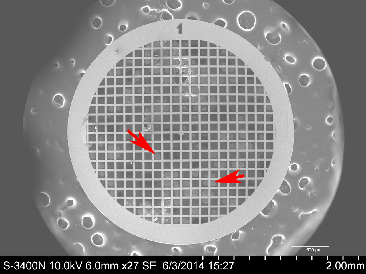
The TEM grids are very small - you have to handle them with fine forceps.  (Notice the torn grid at the bottom of the picture.  I did that picking it up.)  The large, ridged object on the left-hand side of the image above in the tip of my index finger.
Here is an image of the TEM grid in my scanning electron microscope magnified 27x.  The arrows indicate areas where the bacteria have formed on the film between the square supports of the grid. Those spots contain lots of bacteria - way too many.  I imaged in areas of the grid that had the fewest cells.

Micrococcus luteus

Micrococcus luteus is a Gram-positive coccus that is found on human skin as well as in water, dust, and soil.  Micrococcus is generally non-pathogenic, but can cause problems in people with compromised immune systems.  (https://microbewiki.kenyon.edu/index.php/Micrococcus)  This image was made by combining the images from two different detectors on the microscope, the secondary electron detector and the backscatter detector.

Micrococcus luteus [6,020 x]
This is one of the first images I took of M. luteus and I was very pleased with it.  The white dots on the cells I believe to be charging caused by the electron microscope.  These are prokaryotic cells and don't have membrane-bound nuclei.

Another pretty cool thing the SEM can do is mix signals from the two different detectors as different colors.  I chose one detector to be red and the other to be blue.  The result are some very lovely purplish bacteria.

Micrococcus luteus [10,000 x]

Micrococcus luteus [37,000 x]
To get this image I maxed out the voltage on the SEM - 30,000 volts.  Diplococcus is one form M. luteus can take (dipl-  Greek diploos, double; cocc-  Greek kokkos, a kernel or grain.) The "dots" on the surface of the cells are still there, but there was no charging. I have no idea what they are. 
Micrococcus luteus [30,100 x] - Tetrad
Micrococcus luteus often occurs as a tetrad or group of four cells.  
What the heck.  Fun with Photoshop!

Bacillus subtilis

Bacillus subtilis cells are rod-shaped, Gram-positive bacteria that are naturally found in soil and vegetation.  (http://microbewiki.kenyon.edu/index.php/Bacillus_subtilis)

These bacteria gave off electrons at a higher rate and were much easier to image.

Bacillus subtilis [3,730 x]
Bacillus subtilis [6,500 x]
B. subtilis is about 2 microns by 0.7 microns.  This image was made with only the backscatter detector.  Compare to the image below, which is at higher magnification, but of better quality because it is a composite of signals from two detectors.
Bacillus subtilis [11,000 x]
Secondary electrons plus backscatter electrons.

Bacillus subtilis [11,000 x]
Same image as above, but with color mixing.

Bacillus subtilis [27,500 x]
This is my first serious attempt to image bacteria and I learned a lot.  Once they begin teaching microbiology again in the fall semester, I hope to be able to image every bacterium used in the lab. My colleagues are more than happy to let me sneak in and take samples from time to time.

One last note for any students who might be reading.  Notice that scientific names - genus and species - are always italicized and that the genus name is always capitalized while the species name is never capitalized.  After you use a scientific name for the first time in whatever you are writing, it is common and acceptable to shorten the genus to a single letter.  Bacillus subtilis becomes B. subtilis.  Do this and your professors will think you know what you are doing.  Don't do it and they will not smile kindly upon you.

All images in this blog are covered by a Creative Commons Attribution - Non-Commercial 4.0 International License.  Feel free to used, copy, download, or modify the images anyway you like, just please give credit to Eastfield College, Mesquite, TX and don't sell them.

I welcome any comments on this blog.

Murry Gans


Tuesday, May 13, 2014

Electron Microscopy and George Washington


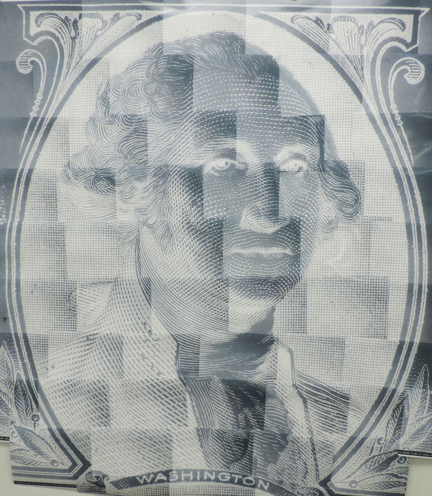
George and me.
Mosaic portrait from one-dollar bill magnified approximately 40 x.
(Microscopy lab coordinator included in picture for scale.)

Full Portrait

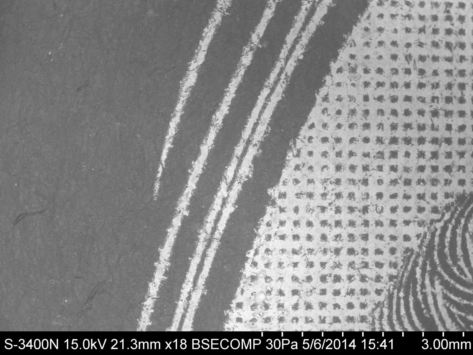
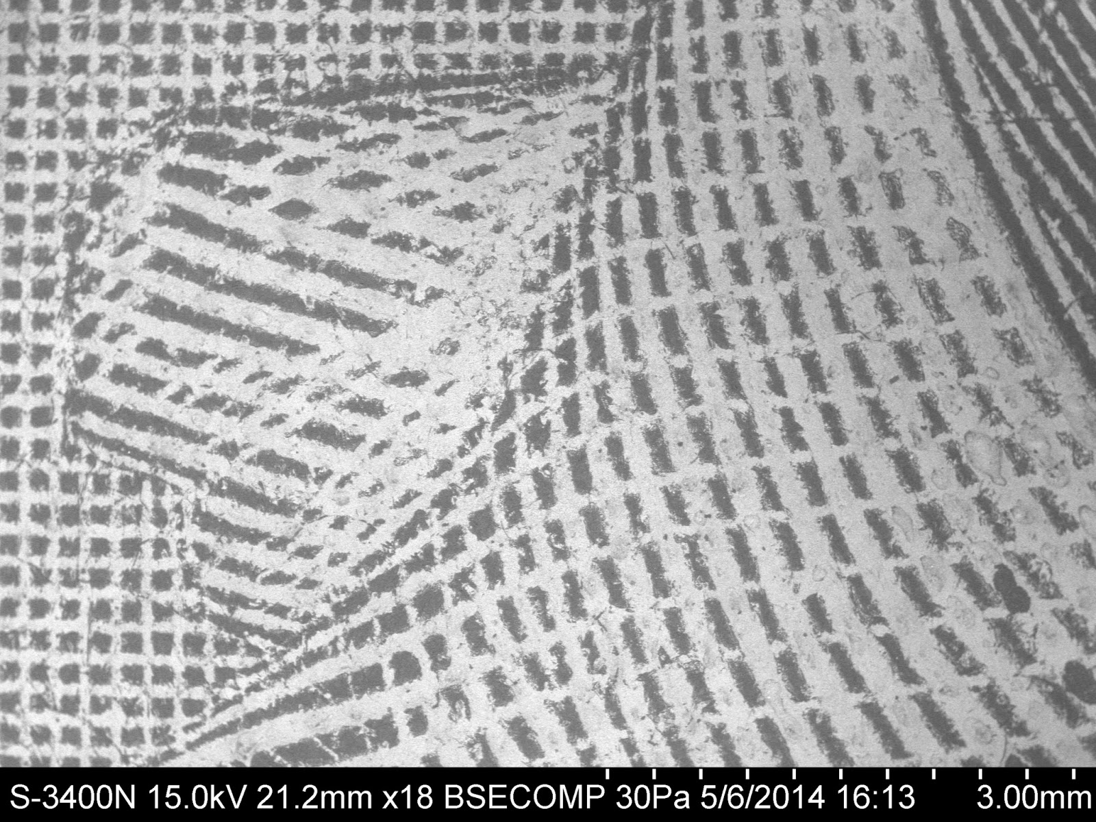
A few months ago I put together a mosaic image of an acorn weevil from eleven overlapping images.  That mosaic, which is about 4 feet long, got a lot of comments so I decided to try something a little different.

When I give presentations to students I like to include images of money because it is so familiar and the ink they use for the intaglio printing on money reflects electrons extremely well. 

I chose the portrait of President Washington because it is relatively small - portraits on the newer fives, tens, and twenties are much larger.  (I also assume the portraits on the fifty and one-hundred dollar bill are also larger, but I can't remember the last time I had one of those.)


The original magnification for each of the fifty-plus images needed to make the mosaic is 18x, which is the smallest I could make the images on my scanning electron microscope.  Once printed, however, the 3 cm wide portrait on the dollar bill became 130 cm wide - a magnification of approximately 40x.  

For those of you here at Eastfield, the mosaic is on the wall outside of the Microscopy Lab - C350. No need to worry if you can't make it to our campus.  Below are all of the images and instructions for making your own mosaic.

Making Your Own Mosaic


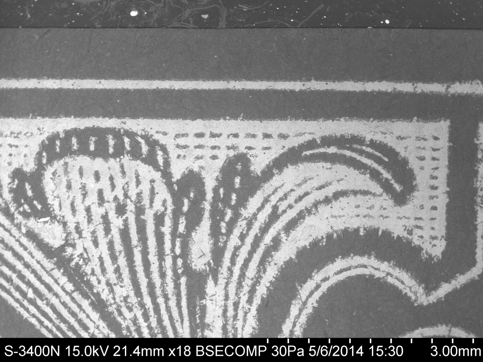
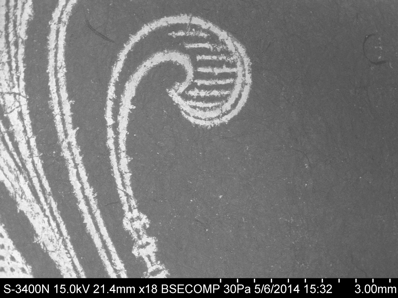
Below are all of the images I used to make this mosaic, grouped by row.  If you would like to make your own just right click on each image and select "Copy Image", then paste it into Photo Shop.  Print them in landscape and check the "Scale to fit media" box.

I began imaging in the upper left hand corner of the portrait making sure to leave an overlap between images so they could be matched up not only with adjacent images, but also those above and below.

I printed each image the size of a full page and then trimmed off the left, right, and top margins.  No need to trim the bottom because the addition of each new row will hide it.  I assembled the first couple of rows on a flat surface and, if I were younger, would probably have put the whole thing together on the floor.  Instead, I hung the assembled rows on the wall with push pins and then added the rest in place.  

Initial assembly was with scotch tape and then reinforced on the back with masking tape.

Important!  Pay attention to two things.  (1)  Make sure that George lines up correctly.  (2)  Make sure that the outside fancy border lines up correctly.  You can fudge a little on the pattern in between.

Row 1 - Left to Right


Row 2 - Left to Right


 Row 3 - Left to Right


Row 4 - Left to Right


Row 5 - Left to Right


Row 6 - Left to Right


Row 7 - Left to Right


Row 8 - Left to Right


Row 9 - Left to Right


Row 10 - Left to Right - These are trimmed off and used to complete the "Washington" lettering and scroll work.


I don't know if anyone is crazy enough to do this (except me) but here you go.  If you put one of these together I would love to see it.  Send me an e-mail.

Murry Gans
Eastfield College