Wednesday, October 17, 2012

A Kidney Stone - OUCH!!


I have done my best to make sure that everyone knows that Eastfield College has a Scanning Electron Microscope that students and faculty can use.  In fact, if you read this blog at all, you know that I say that in every post. (Oh, look. I just did it again!) As a result several students show up to use the SEMs each day and I am beginning to take our portable SEM to area schools. 

One thing that I really didn't expect is people bringing me things to image, and today I was handed my most interesting specimen - and I do mean specimen.  A kidney stone that one of my colleagues passed yesterday.  (I will not tell you who - he or she can tell you if they want to.)

I am told that passing a kidney stone is an extremely painful experience.  The images below really show why that is true.

So brace yourself for an up-close and personal look at a kidney stone.

 Dissecting Scope Image
This is not the entire stone - a small piece broke off

Dissecting Scope Image

SEM Image[20x]

SEM [249x]
Take about death by a 1000 cuts, this view down one edge really shows the angular crystals of calcium and uric acid.

SEM [850x]

SEM [500x]

SEM [208x]
Top view showing not only angular crystals, but also round nodules

SEM [750x]

SEM [250x]

SEM [400x]

SEM [800x]

SEM [2,000x]

SEM [65x]
Interestingly, part of the kidney stone was not made of sharp and nodular crystals.  There was a more solid portion.
SEM [200x]

SEM [561x]
This image, and the one below, show some of the cost of a kidney stone.  The dark patches are tissue from the donor.
SEM [1,600x]

Definitely one of the most unusual and scary things I have imaged.  I don't know about you, but I am going to drink more water!!

See you in the lab.


Friday, October 12, 2012

My Favorite Harbinger of Fall

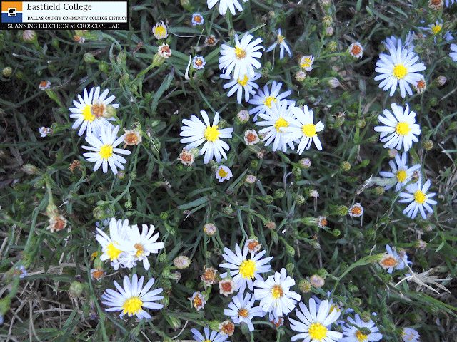
The Late Blooming Aster

(Symphotricum patens - I think)


I love warm weather, but Texas summers are often pretty brutal. I am always delighted when the break in the weather occurs in the fall and our temperatures drop from highs in the 100's to highs in the 80's - often over night. 


When this little aster shows up in my yard and I know that fall is near.  Now that the temperatures have finally changed for the better, these asters are everywhere. 

With flower heads only about 5/8ths of an inch across, these little guys are often over looked, but if you take the time to really look at them, you are in for a treat.  The petals are white to lavender.

 Dissecting Scope Image
This flower head is in a small jar filled with water to keep it turgid. 

Dissecting Scope Image
This is not a simple flower, but a composite flower - made up of several individual flowers.  The flowers in the middle are called disk flowers and have no petals.  The flowers on the outside are called ray flowers and have only a single petal.  If you count the petals you will know how many ray flowers there are.

Dissecting Scope Image
A close up of the disk of this composite flower.  Notice on the image above that 8 of the disk flowers have yet to open.  The little dots in the image are pollen grains.

Disk Flowers - SEM [20x] 
Even though this is only a 20x magnification, the SEM provides excellent depth of field which allows almost the entire flower disk to be in focus.  The flowers in the middle are not open. The flowers farther out have opened and the anthers are protruding.  These will also open up and a stigma will emerge from the center.

 Outer covering of anthers (pollen obvious) - SEM  [120x]

 Anthers - SEM [25x]
On the bottom left of the image you can see a stigma from a ray flower.

 Dissecting Scope Image
Pollen on anther

Composite flowers can be difficult to figure out, so I took the flower head apart to look at the disk and ray flowers.

 Dissecting Scope Image

 Dissecting Scope Image
This is a ray flower with its single petal.  Seed dispersal in this aster is just like a dandelion - once the seed forms in the ovule it will be dispersed by wind.  The pappi in the image will become the parachute that carries the seed.

Dissecting Scope Image

A close up of the stigma of a ray flower.  I am very pleased with this image - you can clearly see the pollen grains on the stigma

But what about those stamen?  They really don't look like the stamen on simple flowers.  In the images below I have used a needle to dissect a disk flower to find the stamen.

 Dissecting Scope Image
Even on this low magnification image you can see that the anthers on the inside of the flower.

 Dissecting Scope Image
An excellent image showing pollen lined up on an anther.

 Dissecting Scope Image
With the disk flower fully open, the stamen are finally revealed.  The anthers are covered in pollen and supported by filaments.

 Tip of unopened stamen - SEM [55x]
The pollen is essentially oozing out of the stamen

Close up of stamen with pollen - SEM [185x]

 Anther with lots of pollen - SEM [62x]

 Filaments that support anthers, Pappi - SEM [71x]

The final two images are of pollen.

 Pollen - SEM [750x]

 Pollen - SEM [1,000x]
Pollen about 25 microns in length - small!

It was a lot of fun to study the composition of this aster using the tools available in the Scanning Electron Microscope Lab here at Eastfield College.  It is amazing what you discover when you are able to see the world in such tiny detail.

The SEM Lab at Eastfield College is open to students and faculty everywhere.  We have two scanning electron microscopes.  Our smaller scope is so simple to use that I can have you capturing images within 15 minutes.  The larger scope provides more flexibility and control, and I am here to help you use it. 

If you are interested in seeing or using our scopes, please contact the lab.

Murry Gans
Scanning Electron Microscope Lab Coordinator
Eastfield College
Mesquite, TX

Thursday, October 4, 2012

It's a bee - no, it's a fly - no, it's a BEE FLY!


The biology professors here at Eastfield College often take their students into the field.  Professor Ron Beecham took a group of students to Gus Engeling Wildlife Management Area last weekend and brought some samples back to the lab for me to image.  One of these is a bee fly - genus Bombylius.

This little guy is easily recognizable as a fly because it possess a single pair of wings and a pair of halteres - reduced wing stubs that are thought to act as counterbalances.  In fact, the order for flies is diptera, which literally means "two wings". ("di-" is pair and "ptera" is wing.)

 Dissecting Scope Image - Ventral View
Note the long tongue

 Dissecting Scope Image - Ventral View
The haltere on the left side of the fly appears as a yellowish blob due to the limited depth of field of the camera on the scope.

Dissecting Scope Image
Dorsal View

As I examined this guy, I noticed a tuft of some "hairs" on the leading edge of the wing near the body.  This tuft if visible in the following two images.

Dissecting Scope Image
Tuft of hairs on right wing, near body.
Dissecting Scope Image
A closer look at tufts of hairs on right wing.

Now for a head-on view of the fly.  The multiple facets of the ommatidia that make up the compound eye are obvious, as are the antennae.

Dissecting Scope Image

Now on to the Scanning Electron Microscope.  Here is pretty much the same pose and you can see the advantages, and single disadvantage, of scanning electron microscopy.  The only disadvantage with the SEM is loss of color since we are using a beam of electrons instead of light.  Not much of a disadvantage at all as far as I am concerned.  One big advantage, which is very obvious when comparing the image above with the one below, is the depth of field.  On the top image I focused on the upper surface of the eyes - the rest of the image is out of focus.  Below you can see that much more of the specimen is in focus.

Head shot
25x


 Head shot
37x
My, what beautiful eyes you have!
 Right eye
85x

Ommatidia of the compound eye - 20 micros across
267x

Antenna
Note the pores
160x

The next series of images are of the haltere, or reduced second pair of wings.


 Haltere
60x


Close-up of haltere - everything has structure
170x

(I realize as I am labeling this image that I missed an opportunity.  Is the structure on the haltere similar to that on the wings of the insect?  A question needing an answer - sounds like an opportunity for someone to do some science.)

As you may have noticed, this little fly has a very long tongue.  Let's see what it looks like

 Tongue
80x
At a higher magnification you can see some pollen grains that are stuck to the tongue.
98x
 Pollen grains and tongue structure
From the scale bar you can see that the pollen grains are between 30 and 35 microns in diameter.
394x

 Tip of the tongue
145x

 Tip of the tongue
447x

 As I was imaging the tongue I noticed some interesting structures on the legs of the fly.  They appear to be covered with scales very similar to those found on butterflies.  This may be normal, but being new to SEM work, it definitely surprised me.


 Tongue in foreground, leg with scales behind
140x
Leg with spines
50x
 Leg joint showing scales and fine structure of spines
190x
Science is all about making observations, asking questions, and then finding out the answers to those questions.  What is the function of scales - on flies and butterflies?  Is it possible they have some aerodynamic effect?
Now a look at the wings. 


 Wing
Note the "hairy" structure on the leading edge of the wing at the top of the image.
27x
 The tufts of "hair" are actually spines and scales.
65x
Scales and spines on leading edge of the wing.
182x


Eastfield College makes it Scanning Electron Microscope lab available to students and faculty at all levels.  If you would like to see or use our SEMs (we have two) please contact me.  Our smaller SEM is portable and can be brought to your school for students to use.  I know, it sounds like a sales pitch, but for high schools, middle schools, and elementary schools there is not cost.

Our lab is also open for visits - bring your classes.  I also Skype!

Murry Gans
Scanning Electron Microscope Lab Coordinator
Eastfield College
Mesquite, TX