This semester I got the chance to teach a microbiology class here at Eastfield College. In lab we are studying fungi so, having some pretty cool microscopes at my disposal, I did some imaging for my class. So this blog will be slightly different since it is intended for my students, but it still has some pretty cool images - even if you aren't in my micro class.
In general, fungi are eukaryotic, unicellular or multicellular, heterotrophic, and have cell walls composed of chitin. (Chitin is also in the exoskeletons of insects and is what goes "crunch" when you step on a bug. The cell walls in plants are composed of cellulose and those in bacteria are composed of peptidoglycan - great test questions, by the way.)
There are lots of different types of fungi and they are everywhere. Watch any local weather report when they do the "pollen count", you will always see fungi. Rag weed may come and go with the season, but fungal spores are always present.
It is really kind of silly to include fungi in a "pollen count" since fungal spores aren't pollen. Pollen is the male gametophyte of plants and ultimately will result in the formation of two sperm cells that will fertilize the egg and the polar nuclei of the ovule to produce the embryo and the endosperm. Fungal spores are haploid cells that are produced by meiosis and are used to disperse the fungus. When they land in a suitable location they germinate and grow.
Yeast - Saccharomyces cerevisiae
The image below was taken by Dr. Tammy Oliver using a Leica microscope with a digital camera attached. This prepared slide shows the general morphology of yeast cells. The original magnification on this image is 1000x using oil immersion.
A prepared slide showing asexual budding. 1000x (oil).
Aspergillus
According to our lab manual, Aspergillus is a Deuteromycota, which means that no sexual reproductive phase as yet been discovered. Of course, that doesn't mean that it doesn't reproduce sexually, just that no one has seen it. (Maybe it is shy.)Aspergillus reproduces asexually by forming conidia (conidium, singular) on conidiaphores. In our prepared slides the conidia are grouped in what appears to be a sporangium, but it isn't, so be careful.
Hyphae are composed of cells that are connected together, end to end. They may be separated from each other by internal cross walls, in which case they are called septate hyphae. If they don't have cross walls they are aseptate hyphae. Even if they have cross walls, those cross walls are very porous allowing the sharing or cytoplasm and organelles, including nuclei. In essense, the hyphae are long continuous cells with multiple nuclei. This condition is called a syncytium.
In the image below you can see the mass of hyphae that make up the mycelium, or body of the fungus. I have also indicated a conidiophore - the filament that holds up the structures that make the conidia spores. Raising the spores up above the mycelium helps them spread.
The image below shows the conidia and the conidiophore.
Dr. Tammy Oliver made the image below from a prepared slide. In this image you can see the conidia and some of the detail of the underlying structures.
Here is a closer look.
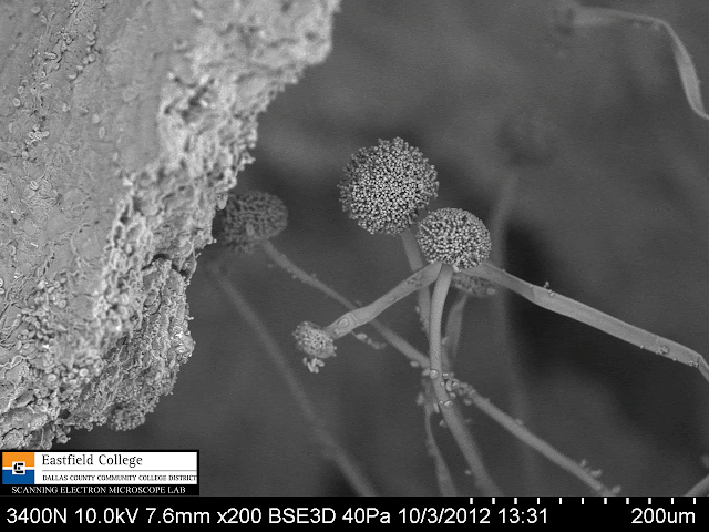
Eastfield has two scanning electron microscopes. We took the smaller, table top SEM into the microbiology lab today to allow the students to see it in action. The images of Aspergillus were taken during lab using a Hitachi TM-1000 SEM.
Aspergillus conidiophore and conidia. [500x]
Hitachi S-3400N [200x]
Hitachi S-3400N [440x]
Hitachi S-3400N [750x]

Conidia [2,010x]
Conidia spores [1,200x] Image made during lab.
Conidia spores [6,000x]
'
One of the cool things about SEMs is that you can make calibrated measurements directly on the image. On the image above you can see that these fungal spores are about 4 microns in diameter.
Rhizopus nigricans
Some time ago I brought a piece of moldy bread into the lab for imaging. This was a specialty bread baked by my local grocery store which meant it didn't have any preservatives added. It molded beautifully.First let's take a look at images made with the digital dissecting scope.
Most people think of moldy bread as pretty disgusting, but once you see it under a microscope the colors and structures are pretty amazing. Unfortunately, I don't have the luxury of a prepared slide to tell me what I am seeing, but I am pretty sure the the black sporangia (spore-forming bodies) are black break mold - Rhizopus nigricans and the white mold in the middle with green under it is Penicillium.
Sporangia of Rhizopus nigricans
Green and white mold of Penicillium
Notice the edge of the white mold just below the yellow You can begin to make out the conidia and conidiaphores of Penicillium.
In the center of the image you can clearly see the white, branching conidiaphores of Penicillium.
More white conidiophores (I seem to be obsessed!!)

Conidia and conidiaphores on moldy bread. The pebbly substrate is bread! [500x]
To make the images below I cut a cross section through the moldy slice of bread. In fact, you can see the voids caused by the production of carbon dioxide during fermentation. The bread is to the left with the mycelium of the fungus above.
Bread and mold [73x]
Bread on bottom left corner. Hyphae and mycelia above. [200x]
Sporangia and sporangiophores of Rhizopus [27x]
Sporangia and sporangiophores [50x]
The funciton of sporangiophores is to get the sporangia up above the surface of the bread so the spores can spread more easily. [50x]

[250x]
Zygospores [2,010 x]
Basidomycota - the mushrooms
These first images were made by a student on the table top SEM.
.gif) This image shows a gill from under the cap of the mushroom. You can clearly see that the basidiospores occur in groups of 4. They extend from a club-shaped basidia which give this groups its name. [200x]
This image shows a gill from under the cap of the mushroom. You can clearly see that the basidiospores occur in groups of 4. They extend from a club-shaped basidia which give this groups its name. [200x]
Basidiospores in groups of 4 [800x]
From a prepared slide - a cross section through the stalk and gills of a Coprinus mushroom.
Prepared slide. Original magnification 1,000x (oil)
Prepared slide. Detail showing basidium and basidiospores. Original magnification 1000x (oil)
WOW. That is a lot of fungi and hopefully the images are interesting and will be useful to my students. This is the first time that I have included images from 4 different microscopes - the TM-1000 SEM, the S-3400N SEM, a digital dissecting scope, and a digital light microscope, which I think is pretty neat.
As always, I would appreciate any feedback on the blog.
Eastfield College Microscopy Lab
Eastfield College
Mesquite, TX


.gif)


.gif)
.gif)


.gif)


That was very helpful professor, it is amazing that some of those pictures are of mold, and fungus.....Have a great night and thanks for the pictures it will be most helpful on the next test...
ReplyDeleteCourtney
Is there a procedure I can follow to view basidium and their spores?
ReplyDeleteEmily,
ReplyDeleteThe slide that I used to make the light microscope images of the basidium was a prepared slide from the lab. It looks like the folks who made the slide used a microtome to make a thin cross-section through the gill of a mushroom. I have to admit that I haven't made this type of slide myself, but if I were to try it I would use a new razor blade to make my thin cross-section and then make a standard wet-mount slide. You might even add a little methylene blue to stain the calls. I have found that in most cases, if you are patient and look around on the slide, you will get lucky. This seems to work well for me in the past. Let me know how it works out.
My electron microscopy images were made with no sample preparation at all, other than mounting a gill. Ideally, I would have used a critical point drier to keep the spore structure from collapsing under the vacuum of the SEM and would have sputter coated the sample with gold/palladium. Unfortunately, the college doesn't yet have these machines but I am eternally hopeful that one day we will.
I have seen students in my lab do pretty cool things just by trying, so don't be afraid to give it a try.
Good day Professor,
ReplyDeleteMy son (12 years) is doing a science project on fungi at school and we by pure luck managed to land on your blog. I have never seen my son so ecstatic about fungi or anything else. I suppose I should not be surprised as his goal and dream in life is to go and work at NASA one day.
Thank you for making his day :-) , the images are absolutely awesome. He will be drawing his own sketches and I can not even start to tell you how helpful your slides are going to be! (He is insisting on using the proper scientific method for his project, so we have a long stretch in front of us!)
Thanking you from a sunny South-Africa.
Wilana and Rogan Olivier
Dear Professor,
ReplyDeleteI would like to study the effect of silver nanoparticle on Fusarium solani. For the same I want to perform SEM for the same. I never worked on fungi. Can you please guide me how to proceed for this experiment. I read about fixation and dehydration. I would like to know how to grow fungi in nanoparticle sample. Should I grow it in liquid media on cover slip? or to grow in PDA agar plate (added nanoparticle). How to remove agar for final observation?
Please reply.
Thanks
Sarika Pawar
Hi Murray, Let me come straight out with it - I'm a layman dinosaur - albeit one that's done a lot of self educating! I've always been very interested in EVERYTHING! And not just for an overview but right down to the nitty-gritty - I just love the InterWeb!! As it brings folk like you and your work into my life at a few clicks of a mouse ( in my younger days they only squeaked!)
ReplyDeleteThough I studied Biology in U.K. senior school and went on to do a lot of practical Horticulture, much of what the younger generations read up as background had yet to be discovered - my Biology text book (1925 vintage) has about 200 words to say about photosynthesis - some of the chemistry was about right but they had no idea of electron excitation, ATP or NADP etc.
Since retiring ( though I'm so busy, I don't know how I fitted work in!) from what's called Landscape Gardening over here (earthmoving for you I guess) I've been doing more research into the latest discoveries - especially if they impinge on any of my pet theories!
I'm particularly interested in sex! (Hmmm.. aren't we all?) Ah... but in this case - it's origins in early life and its development in plants. Having prokaryotes going about dividing is a long way from flowering plants and the resulting seed pods - needs some explaining!
I've lucky enough to find quite a few academics around the world that were willing to answer a few "stoopid" questions. I'd like to ask your opinion of ancient symbiosis - it's almost certainly responsible for the inclusion of organelles in the cell and maybe more than once and as there's a lot of discussion about meiosis being a response to parasitic infection - I thought of micrograms of germinating spores. However; the big problem in my mind to all these periodic additions to cells - is the transcription of the "improvement" into the existing RNA/DNA without the various repair mechanisms getting rid of it.
Any thoughts? And if any of your followers want to jump in - be my guest! Many thanks
You’ve got some interesting points in this article. I would have never considered any of these if I didn’t come across this. Thanks!. educational technology
ReplyDeleteThis is great professor. This is really helped in studying organisms in my college Biology Lab class where we unforuntately cannot view specimens due to online classes. I do appreciate your efforts in taking such incredible photos of these specimens. They turned out really well!
ReplyDelete