Eastfield College has some beautiful courtyards with lots of shade and benches. Even in our extremely hot Texas summers I can always find a place to sit and relax for a few minutes.
The other day I was sitting on one of my favorite benches when I noticed that the rosemary in the planter next to me was covered in spider webs. I am a good enough arachnologist to realize that where there are spider webs there are spiders. I took a quick look but couldn't find the little creatures so I clipped off a sprig of rosemary and took it back to my scopes in the SEM Lab.


Once I got the sprig of rosemary back to the lab I put it on the dissecting scope and began my search. Within a few minutes I found the little guy.


Another very small Theriidian spider. (You can see another species of this same family of spiders in a previous blog.) These spiders are commonly referred to as cobweb spiders. Black widow spiders belong to the same family.
By this time you can imagine that my lab and hands are smelling very strongly of rosemary. All of that aromatic oil must be stored in glands on the leaf.

A look at the top surface of the leaf shows the expected oil glands. If you look carefully at the image above you can see there are two different sizes of glands. Do they contain different oils? Sounds like a good question for someone with a gas chromatograph.
As observant as I try to be I am a bit embarrassed to admit that I had never looked closely at the leaf on the rosemary bush - and I have one in my own backyard.
What I didn't realize is that the top surface of the leaf curls around to the bottom of the leaf.


The images above illustrates a problem - a limited depth of field. How to solve that problem - don't use light - use electrons in the SEM.
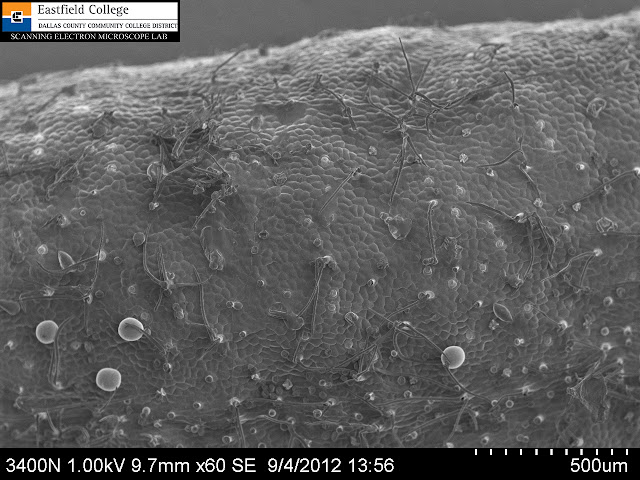
Top surface of rosemary leaf. [60x]
The image above shows the epidermal cells of the leaf as well as the two different sizes of oil glands.

Lower surface of the rosemary leaf [24x]
Note how all levels of the leaf are now in focus.

Lower surface of rosemary leaf [70x]
This image shows the midrib with smaller oil glands. You can also see some spider web stretching diagonally across the upper half of the picture.

Spider web [99x]
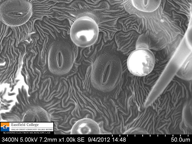
Stomata and oil glands [1,000x)

Stoma portrait [2,490x]
The high vacuum and electron beam of the SEM dry out specimens. This stoma was open when I first saw it, but quickly closed as the guard cells lost their turgor.
As I use the instruments in the SEM Lab at Eastfield College I am discovering that even the most common organisms have some pretty fantastic morphology under high magnification.
The imaging instruments at Eastfield College are available for research to students and faculty everywhere. Want to get a better look at your world - give us a call.
Murry Gans
Scanning Electron Microscope Lab Coordinator
Eastfield College
Mesquite, TX
972-860-7267

No comments:
Post a Comment