Friday, December 14, 2012
Thursday, December 6, 2012
A Magnificent Bug - Part II
Yesterday's blog post took a look at our not-so-little friend shown below using a camera and dissecting scope. Now it is time to take a look with the scanning electron microscope.
Say hello to my little friend.
Here is the SEM I used to make these images - the Hitachi S-3400N.
[6x] SEM Image
One of the things that amazed me when I saw this insect with the SEM is how toughly built it is - lots of strong skeletal elements, including spikes. Notice also the long, piercing mouthpart tucked under the body. Also visible on the abdomen to the far right of the image are two of the spiracles the bug uses for breathing.
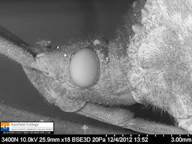
[18x] SEM Image
A closer look at the compound eye of the insect. The smaller bump just above the compound eye is a simple eye. This is actually bright red. (See yesterday's images of this bug.)
[7x] SEM Image
A head-on image. Notice the raised horn between the antennae and the bed of small, chitinous spikes across the surface of the thorax behind the head.
[21x] SEM Image
Good to see you! A closer look at the face and the horn.
[35x] SEM Image
The bright white specks in the image are actually dirt, dust, or possibly plaster flakes from the insect killing jar.
[18x] SEM Image
For this image I placed the insect on its back. The two almost vertical structures to the left and right of the image are front legs. Notice the strange structure of the mouthparts. The next two images zoom in on parts of these.
[55x] SEM Image
Top of the mouthpart - where it connects to the head.
[150x] SEM Image
OK. I will admit that I have no idea why this structure looks like this, but of course that is how science works. You find something you didn't know about, start asking questions, and then work at finding the answers.
[17x] SEM Image
A dorsal view - look straight down on the insect. In this image you can see the two compound eyes and the two simple eyes next to them. Besides lots of hairs, you can also see a pair of spikes in the center of the thorax and along both edges. This species must have a pretty tough life to need such armor. Something else to find out about.
[158x] SEM Image
A close up of a couple of small spikes that cover the dorsal surface of the thorax. You can see scratch marks on the top spike.
[190x] SEM Image
This is one of the spikes that extend from the margin of the thorax. From the scale bar on the image you can tell that the spike is approximately 300 microns long - about 1/3 of a millimeter (1/10 of an inch).
[11x] SEM Image
This image shows the side of the abdomen near its connection with the thorax. The edges of the wings are visible at the top. Here I discovered two interesting structures - most likely sensory organs. (I am going to have to go to the library and hit the Internet - lots to find out here.)
[50x] SEM Image
A closer look at the upper sensory structure - about 300 microns (1/10 of an inch) at its widest point.
[170x] SEM Image
A fine mesh of hairs covers this opening. Why?
[88x] SEM Image
This is the lower opening - this could be a spiracle of breathing. Not the rough structure of the insect exoskeleton.
[20x] SEM Image
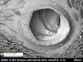
As noted in the previous blog, insects breath via a series of tubes called trachea, that permeate their bodies. These trachea open directly to the atmosphere through holes called spiracles. This image shows two of those spiracles on the side of the abdomen.
[142x] SEM Image
Spiracle opening
[200x] SEM Image
Spiracle opening. Note the internal structure.
[85x] SEM Image
This image shows the tarsal claws on one of the legs. The two sac-like structures to the right of the claws are sense organs. Also, notice the hairs on the part of the leg shown at the bottom of the image.
Having worked in the SEM lab for about 6 month now I realize that even though the SEM will let me greatly magnify specimens, sometime lower magnifications can show the most. The highest magnification on this series of images is only 200x - well within the range of a light microscope. So why use the SEM - because of its ability to show excellent depth of field and razor sharp focus.
As always, you are invited to visit the SEM Lab at Eastfield College, either in person or electronically. I look forward to hearing from you.
Murry Gans
Scanning Electron Microscope Lab Coordinator
Eastfield College
Wednesday, December 5, 2012
A Magnificent Bug - Part 1
Two days ago biology Professor Ron Beecham walked into my office with a truly magnificent specimen - a Leaf-footed bug. For once I get to use the term "bug" as it should be used, because this insect belongs to Order Hemiptera - the true bugs. Technically, this is the only order of insects that should be called bugs.
This was such an interesting specimen that I took too many images to post in one blog, so I am going to post it in two parts.
Hemiptera literally means "half winged." According to A Source-Book of Biological Names and Terms by Edmund Jaeger (copyright 1972) the prefix "hemi-" means half, and "pter" means wing. [By the way, most of the order names for insects contain "-ptera", which makes lots of them easy to remember.]
The "half-wing" designation is obvious in the picture below - the forward pair of wings have a thicken base and are more membrane-like toward the tip. This makes the wings look like they are half covered.
This bug belongs to family Coreidae - the leaf-footed bugs. According to Peterson's Field guide to Insects "This is a large group, and most of its members are relatively large bugs. . . . Some are plant feeders and others are predaceous. . . . Coreids often give off an unpleasant odor when handled."
I can definitely attest to that last statement!!
The following images were takien on a dissecting scope with a digital camera attached.
Here is a closer look at the characteristic that places this insect in Order Hemiptera
Here you can see the flattened extension at the distal end of the hind legs. Why is it there? What comes to mind is that it might be used by the insect to control its flight. That would be an interesting premise to test.
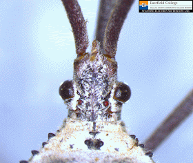
A close up of the dorsal side of the head. The two compound eyes are obvious as are the two red simple eyes.
My, what pretty red eyes you have (or not).
This frontal view shows the spiky armor and, at the front of the head, a horn.
Flip the bug over on its back an you can see its piercing weapon - great for sucking sap from plants and drilling into other insects. What you see here is actually the sheath the encloses the piercing stylets. The horn on the head is also visible here.
A closer look at the ventral side of the head.
This image shows the left side of the abdomen. Insects do not have lungs like we do. Instead they have a series of tubules, tracheae, that permeate all parts of their bodies and are open to the outside atmosphere via openings called spiracles, indicated on this image by the red arrows.Below is a mosaic of several images pieced together to show the entire insect. It was way too big to see at one time with the scope.
Part II will show scanning electronmicrographs of this same insect.
As always, you are invited to come by or contact the SEM Lab at Eastfield College. We support STEM research at all levels.
Murry Gans
SEM Lab
Eastfield College
Monday, December 3, 2012
Fall Colors - On a Small Scale
Eastfield College is located in north central Texas, which is not exactly known for its fall colors. We have lots of yellows and browns (aka - dead leaves). In fact, as I sit here this late afternoon and look out my office window, the colors aren't exactly inspiring.
As I was walking on campus yesterday afternoon, however, I came across a Sweet Gum tree in one of our courtyards. This time the colors did catch my attention. (For those of you who work or study on campus, this tree is located in the courtyard formed by the S, C, and N Buildings.)
The following series of pictures were made with my little point and shoot digital camera.
Looking East - the S Building in the background
Looking South - C Building in the background
In this image you can see the spiky fruit of the Sweet Gum tree
I was quite pleased that while I was taking these pictures the people who walked by actually looked up! One of those people was a campus police officer who politely told me to stop walking around in the flower beds.
Of course they pay me the big bucks (I wish) to let people see the world from a different perspective, so instead of looking up, let's look down - specifically at the fallen leaves.
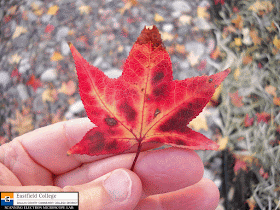
I didn't arrange the leaves in this shot. I just looked down where I was standing and took the picture.
We may not have lots of trees with amazing fall colors, but what if we just looked at a single leaf - a change of perspective.
(No professional hand models were used for these images - those stubby fingers are mine.)
So now back to the lab for a closer look at different areas of single leaves. The images below were made with the lab's digital dissecting scope. I will admit that once I started looking closely at each leaf, I kept finding more and more areas to image. I went a little wild so there are lots of images below. I will let them speak for themselves.
Pretty and cool! I also took about 30 images with the scanning electron microscope, but black and white images would seem out of place here.
Happy Fall!
Scanning Electron Microscope Lab
Eastfield College


